The University of South Florida’s Center for Advanced Medical Learning and Simulation (CAMLS), part of the Tampa Medical & Research District (TMRD), has become the first institution in Florida -- and third in the world -- to install GE HealthCare’s (GEHC’s) Allia Moveo image-guided therapy system.
As an additional milestone, CAMLS, which is one of the world’s largest free-standing simulation facilities dedicated exclusively to healthcare training, will also become the first simulation center globally to integrate Allia Moveo into its education and simulation programs for students, faculty, and practicing clinicians, CAMLS said.
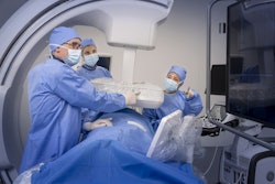
The addition of the Allia Moveo system will allow multidisciplinary teams to gain hands-on experience with technology used in complex minimally invasive procedures, CAMLS and GEHC said.
The system’s features include a compact, cable-free C-arm enabling full movement and clear patient access; a wide-bore C-arm designed to accommodate a diverse patient population and enable conebeam CT imaging; and AI-powered workflow tools to streamline procedures and help deliver more personalized patient care.
A second Allia Moveo system is scheduled to be installed in the hybrid operating room suite at Tampa General Hospital for clinical use later in 2026, CAMLS and GEHC added.


![Images show the pectoralis muscles of a healthy male individual who never smoked (age, 66 years; height, 178 cm; body mass index [BMI, calculated as weight in kilograms divided by height in meters squared], 28.4; number of cigarette pack-years, 0; forced expiratory volume in 1 second [FEV1], 97.6% predicted; FEV1: forced vital capacity [FVC] ratio, 0.71; pectoralis muscle area [PMA], 59.4 cm2; pectoralis muscle volume [PMV], 764 cm3) and a male individual with a smoking history and chronic obstructive pulmonary disorder (COPD) (age, 66 years; height, 178 cm; BMI, 27.5; number of cigarette pack-years, 43.2, FEV1, 48% predicted; FEV1:FVC, 0.56; PMA, 35 cm2; PMV, 480.8 cm3) from the Canadian Cohort Obstructive Lung Disease (i.e., CanCOLD) study. The CT image is shown in the axial plane. The PMV is automatically extracted using the developed deep learning model and overlayed onto the lungs for visual clarity.](https://img.auntminnie.com/mindful/smg/workspaces/default/uploads/2026/03/genkin.25LqljVF0y.jpg?auto=format%2Ccompress&crop=focalpoint&fit=crop&h=100&q=70&w=100)


![Images show the pectoralis muscles of a healthy male individual who never smoked (age, 66 years; height, 178 cm; body mass index [BMI, calculated as weight in kilograms divided by height in meters squared], 28.4; number of cigarette pack-years, 0; forced expiratory volume in 1 second [FEV1], 97.6% predicted; FEV1: forced vital capacity [FVC] ratio, 0.71; pectoralis muscle area [PMA], 59.4 cm2; pectoralis muscle volume [PMV], 764 cm3) and a male individual with a smoking history and chronic obstructive pulmonary disorder (COPD) (age, 66 years; height, 178 cm; BMI, 27.5; number of cigarette pack-years, 43.2, FEV1, 48% predicted; FEV1:FVC, 0.56; PMA, 35 cm2; PMV, 480.8 cm3) from the Canadian Cohort Obstructive Lung Disease (i.e., CanCOLD) study. The CT image is shown in the axial plane. The PMV is automatically extracted using the developed deep learning model and overlayed onto the lungs for visual clarity.](https://img.auntminnie.com/mindful/smg/workspaces/default/uploads/2026/03/genkin.25LqljVF0y.jpg?auto=format%2Ccompress&crop=focalpoint&fit=crop&h=112&q=70&w=112)


