Routine CT scans used to plan radiotherapy for breast cancer can identify coronary artery calcium and detect risk of heart disease without additional imaging, radiation, or cost, according to a study showing patients with extensive calcification had significantly higher rates of ischemic heart disease during follow-up.
- Study findings: Among 207 patients aged 65+, all six cases of ischemic heart disease occurred in the 58 patients with extensive coronary artery calcification (10.3% incidence rate).
- Zero additional cost: Coronary artery calcium assessment requires no extra imaging, radiation exposure, or financial investment beyond existing radiotherapy planning CT scans.
- Missed opportunities: Only 12.5% of radiation oncologists routinely document coronary artery calcium, though 78.1% would change practice if clinical value were established.
- Hidden risk detection: Four of six heart disease events occurred in patients with no known cardiac history, demonstrating the screening value for asymptomatic disease.
- Actionable strategy: Incorporating structured coronary artery calcium assessment into routine CT reporting could improve cardiovascular risk stratification for breast cancer survivors.
CT scans used to plan radiotherapy for women with breast cancer provide an opportunity to screen for risk of heart disease, according to a study published August 17 in Practical Radiation Oncology.
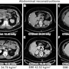
A team from Tel Aviv Medical Center in Israel calculated coronary artery calcium (CAC) scores from routine simulation CT scans from 207 older patients undergoing adjuvant radiotherapy for early-stage breast cancer, with findings suggesting the approach could support cardiovascular risk stratification, according to the group.
“Radiotherapy simulation CT provides a unique and underutilized opportunity to detect subclinical coronary atherosclerosis without additional imaging, radiation exposure, or cost,” noted lead author Inbal Golomb, MD, and colleagues.
As survival after early-stage breast cancer improves, cardiovascular disease has emerged as an increasingly prominent competing risk in older women, the authors wrote. CAC, a validated marker of coronary atherosclerosis, is frequently visible on non-contrast chest CT, and radiotherapy simulation CT offers an underutilized opportunity to detect it without additional cost or radiation exposure, they added.
To explore the strategy, the researchers retrospectively enrolled 207 consecutive patients aged 65 or older between 2007 and 2008, with follow-up through January 2026. Simulation CT scans were visually graded by two radiation oncologists on a three-level CAC score: 0 (no calcification), 1 (mild focal), and 2 (extensive). The primary endpoint was incident ischemic heart disease (IHD) during follow-up.
According to the results, over a mean follow-up of 6.6 years, six patients (2.9%) developed IHD, all among 58 patients with extensive calcification (10.3%). There were zero events among 149 patients with absent or mild scores (OR 22.8, p = 0.0008). Notably, four of the six events occurred in patients with no known cardiac history, the researchers reported.
In secondary results, a survey of 32 radiation oncologists found only 12.5% routinely document CAC on simulation CT scans, while 78.1% indicated a willingness to modify practice if clinical relevance were established.
“For many patients with early breast cancer, simulation CT may represent the only thoracic imaging obtained during oncologic care, and therefore the only opportunity to identify incidental CAC,” the researchers wrote.
Incorporating structured CAC assessment into routine reporting may represent a simple, cost-neutral strategy to improve cardiovascular risk stratification and support preventive care in breast cancer survivors, the group concluded.
The full study is available here.