Deep-learning models used with preoperative CT images can predict the likelihood of malignant and aggressive pathology of incidentally discovered renal masses, researchers have reported.
The study results could help clinicians better diagnose and treat these unexpected findings, wrote a team led by Ying Xiong, PhD, of Fudan University in Shanghai, China. Xiong's and colleagues' work was published February 7 in Nature Communications.
"Under most circumstances, the decision between active surveillance, percutaneous ablation, and surgical resection is made without a reliable pathologic diagnosis," the group explained. "Thus, there is an urgent need to improve the noninvasive diagnosis of benign renal masses and differentiate aggressive tumors (prompting treatment) from indolent tumors (allowing ablation or deferred treatment)."
The increased use of cross-sectional imaging modalities such as CT has boosted discovery of incidental kidney lesions -- which can lead to more surgeries and ablations, the group explained. But most of these lesions don't seem to translate into higher mortality rates, which suggests overdiagnosis. That's why it's crucial to assess incidental kidney masses.
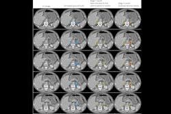
Xiong's group explored whether using deep-learning models with CT could help clinicians categorize these incidental renal masses via a study that included 13,261 preoperative CT volumes taken from 4,557 patients. The team developed two multiphase convolutional neural networks, one to predict malignancy of any identified renal masses and another to predict aggressiveness.
The researchers reported the following:
- Model 1, designed to predict the malignancy of renal masses, achieved an area under the curve (AUC) of 0.87, besting the average performance of seven radiologist readers.
- Model 2, designed to differentiate between aggressive and indolent tumors, achieved an AUC of 0.78.
Both models outperformed a corresponding radiomics model the team developed and a nephrometry score nomogram (a tool that uses the RENAL Nephrometry Score to predict the likelihood of a kidney mass being malignant or high-grade), according to Xiong and colleagues.
The investigators also noted that the deep-learning tool had a positive effect on the radiologists' reading performance -- a finding that underscores the tool's complementary nature.
"With the assistance of the deep-learning model, the diagnostic accuracy of the radiologists significantly improved," the authors wrote. "We observed that seven radiologists displayed a higher degree of diagnostic consistency amongst themselves when compared to the deep-learning model. This suggests that the deep-learning model and the radiologists may be approaching the classification of renal masses from different yet complementary perspectives."
The complete study can be found here.