Photon-counting CT (PCCT) is "the first new quantum leap in CT technology in a decade," according to a presentation delivered September 6 at the International Society of Computed Tomography (ISCT) annual meeting in San Diego.
The technology's forerunners are dual-source, dual-energy CT, but it expands the modality's capability by adding spectral imaging and ultra-high resolution to the tool kit, presenter Joseph Schoepf, MD, of the Medical University of South Carolina, told session attendees.
Schoepf offered a primer regarding what PCCT technology adds to cardiac imaging:
- PCCT's calcium scoring performance correlates with conventional CT, but its measurements are more accurate, according to Schoepf; the same is true for stenosis measurements.
- PCCT represents an evolution of temporal resolution for spectral cardiac CT. Schoepf noted that in 2005, first generation dual-source CT had temporal resolution of 83 milliseconds; in 2021, PCCT showed temporal resolution of 66 milliseconds (lower values translate to better resolution).
| Development of temporal resolution for spectral CT compared with DSCT | ||||
| Measure | 2005, first generation DSCT | 2009, second generation DSCT | 2014, third generation DSCT | 2021, PCCT |
| Temporal resolution in milliseconds (ms) | 83 ms | 75 ms | 66 ms | 66 ms |
"[PCCT offers] spectral data without temporal resolution penalty," he noted.
Schoepf also highlighted that PCCT improves the image quality of coronary CT angiography, particularly in obese patients, and removes calcium from images of the heart -- allowing clinicians to evaluate stenosis severity more precisely. Finally, he noted that PCCT not only overcomes technical limitations -- so that speed and spectral acquisition are "no longer mutually exclusive -- but it also overcomes longstanding cardiac limitations such as dealing with extensive calcifications, imaging stents, and characterizing the myocardium.
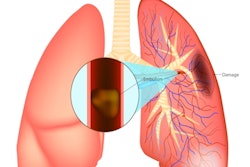
In a presentation delivered during the same session, Philippe Douek, MD, of Lyon University in France also sang PCCT's praises -- in particular for its use in thoracic imaging, with applications ranging from chronic obstructive pulmonary disease, lung cancer screening, characterizing lung fibrosis, and identifying pulmonary embolism and chronic pulmonary hypertension.
"[PCCT's] benefits include ultra-high spatial resolution of the lung parenchyma using small detector pixels, higher matrix size, thinner slice thickness -- as low as 0.15 mm -- and higher frequency filters," he said.
The technology offers improved visualization of small and distal vessels, and lung nodule image quality and conspicuity, Douek said. He presented data that compared image quality between standard dose, conventional CT to standard and low-dose spectral PCCT using a five-point scale (with 1 equal to poor and 5 equal to excellent):
| Spectral PCCT performance for lung imaging compared with conventional CT | ||
| Measure | Conventional CT | Spectral PCCT |
| Overall image quality | 3 | 4 |
| Noise image quality | 3.7 | 5 |
| Distal airways | ||
| Conspicuity | 3 | 5 |
| Sharpness | 2 | 4.3 |
| Distal vessels | ||
| Conspicuity | 2.5 | 5 |
| Sharpness | 2.3 | 5 |
| Proximal bronchial wall | ||
| Conspicuity | 2.7 | 5 |
| Sharpness | 2 | 4.3 |
Spectral PCCT is a game-changer for both clinicians and patients, Douek concluded.
"SPCCT will change the standard of care of thoracic diseases," he said.