
In 93% of patients hospitalized for moderately severe COVID-19, lung abnormalities resolve by their 12-month CT follow-up exam, according to a study published May 10 in Radiology.
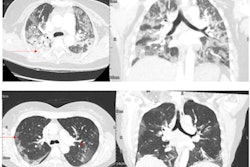
In 78 of 84 patients hospitalized for COVID-19 between March 2020 and July 2021, chest CT scans at 12 months showed complete resolution of lung abnormalities such as ground-glass opacities, consolidation, bronchiectasis/bronchiolectasis, and reticulation, wrote a team led by Dr. Marialuisa Bocchino of Frederico II University of Naples.
"Our results show that residual lung abnormalities on CT are minimal at one year in patients who experienced moderate COVID-19 pneumonia," the group concluded.


![Images show the pectoralis muscles of a healthy male individual who never smoked (age, 66 years; height, 178 cm; body mass index [BMI, calculated as weight in kilograms divided by height in meters squared], 28.4; number of cigarette pack-years, 0; forced expiratory volume in 1 second [FEV1], 97.6% predicted; FEV1: forced vital capacity [FVC] ratio, 0.71; pectoralis muscle area [PMA], 59.4 cm2; pectoralis muscle volume [PMV], 764 cm3) and a male individual with a smoking history and chronic obstructive pulmonary disorder (COPD) (age, 66 years; height, 178 cm; BMI, 27.5; number of cigarette pack-years, 43.2, FEV1, 48% predicted; FEV1:FVC, 0.56; PMA, 35 cm2; PMV, 480.8 cm3) from the Canadian Cohort Obstructive Lung Disease (i.e., CanCOLD) study. The CT image is shown in the axial plane. The PMV is automatically extracted using the developed deep learning model and overlayed onto the lungs for visual clarity.](https://img.auntminnie.com/mindful/smg/workspaces/default/uploads/2026/03/genkin.25LqljVF0y.jpg?auto=format%2Ccompress&crop=focalpoint&fit=crop&h=112&q=70&w=112)


