
Recreational and occupational divers should undergo chest CT three months after recovery from COVID-19 before returning to diving, according to research dated March 31 and published online in Diving and Hyperbaric Medicine.
COVID-19 has been shown to have long-term pulmonary and cardiovascular effects that aren't necessarily linked to disease severity, wrote a team led by Dr. Bengusu Mirasoglu of Istanbul University in Turkey. These effects could make divers vulnerable to diving accidents, which is why it's important to assess their health after recovery from COVID-19 before they return to diving.
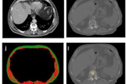
Mirasoglu and colleagues conducted a study of 43 divers who were assessed for fitness to dive with lung CT scans. The team took participants' COVID-19 history into consideration.
At the time of COVID-19 diagnosis, 68.2% of patients had at least one lung lesion; within the first three months after diagnosis, 73.3% had at least one lesion; and after three months, 19.2% had at least one lesion. The most common findings on CT were ground-glass opacities and fibrosis
Of the 43 divers, 13 were kept from returning to diving due to persistent COVID-19-related lung effects.
"Divers who recover from COVID-19 should undergo fitness to dive assessments before resuming diving," the group concluded. "A chest CT performed at least three months after diagnosis may be suggested."


![Images show the pectoralis muscles of a healthy male individual who never smoked (age, 66 years; height, 178 cm; body mass index [BMI, calculated as weight in kilograms divided by height in meters squared], 28.4; number of cigarette pack-years, 0; forced expiratory volume in 1 second [FEV1], 97.6% predicted; FEV1: forced vital capacity [FVC] ratio, 0.71; pectoralis muscle area [PMA], 59.4 cm2; pectoralis muscle volume [PMV], 764 cm3) and a male individual with a smoking history and chronic obstructive pulmonary disorder (COPD) (age, 66 years; height, 178 cm; BMI, 27.5; number of cigarette pack-years, 43.2, FEV1, 48% predicted; FEV1:FVC, 0.56; PMA, 35 cm2; PMV, 480.8 cm3) from the Canadian Cohort Obstructive Lung Disease (i.e., CanCOLD) study. The CT image is shown in the axial plane. The PMV is automatically extracted using the developed deep learning model and overlayed onto the lungs for visual clarity.](https://img.auntminnie.com/mindful/smg/workspaces/default/uploads/2026/03/genkin.25LqljVF0y.jpg?auto=format%2Ccompress&crop=focalpoint&fit=crop&h=100&q=70&w=100)


![Images show the pectoralis muscles of a healthy male individual who never smoked (age, 66 years; height, 178 cm; body mass index [BMI, calculated as weight in kilograms divided by height in meters squared], 28.4; number of cigarette pack-years, 0; forced expiratory volume in 1 second [FEV1], 97.6% predicted; FEV1: forced vital capacity [FVC] ratio, 0.71; pectoralis muscle area [PMA], 59.4 cm2; pectoralis muscle volume [PMV], 764 cm3) and a male individual with a smoking history and chronic obstructive pulmonary disorder (COPD) (age, 66 years; height, 178 cm; BMI, 27.5; number of cigarette pack-years, 43.2, FEV1, 48% predicted; FEV1:FVC, 0.56; PMA, 35 cm2; PMV, 480.8 cm3) from the Canadian Cohort Obstructive Lung Disease (i.e., CanCOLD) study. The CT image is shown in the axial plane. The PMV is automatically extracted using the developed deep learning model and overlayed onto the lungs for visual clarity.](https://img.auntminnie.com/mindful/smg/workspaces/default/uploads/2026/03/genkin.25LqljVF0y.jpg?auto=format%2Ccompress&crop=focalpoint&fit=crop&h=112&q=70&w=112)


