The first set of images of patients with COVID-19 has gone live in the Medical Imaging and Data Resource Center (MIDRC), a collaboration of medical imaging organizations funded by the U.S. National Institute of Biomedical Imaging and Bioengineering (NIBIB).
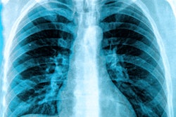
The MIDRC has published an initial set of anonymized images of COVID-19-positive chest CT scans and plans to include x-rays as well. The center seeks to have the largest open database of anonymized COVID-19 medical images, along with associated clinical data, in the world, the MIDRC said.
Freely available, the imaging data may be used for research, education, developing artificial intelligence (AI) software, deep-learning tools, and to serve as a general reference to clinically recognize COVID-19.
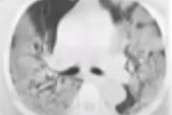
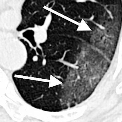
 CT scan of lungs of COVID-19 patient with areas described by radiologists as resembling grains of ground glass. Image courtesy of RSNA.
The RSNA led the charge surrounding COVID-19 images and released the initial set of such images through its RSNA International COVID-19 Open Radiology Database (RICORD). The society is currently processing additional contributions, and it has also partnered with the Society of Thoracic Radiology to source 120 CT scans from four international sites.