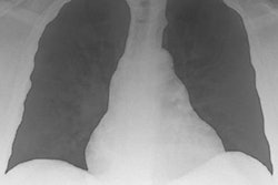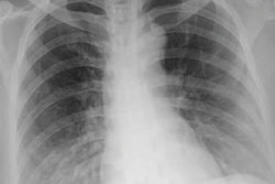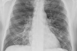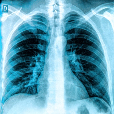
Chest x-rays are effective every three or four days rather than daily for monitoring disease progression in hospitalized COVID-19 patients, according to a study published in the March issue of the Journal of the College of Physicians and Surgeons Pakistan.
An analysis of the use of chest x-ray in patients performed at admission, peak day of findings, and discharge revealed a short plateau phase in findings, followed by improvement at 15 days after initial symptoms. The result suggests there is no need for daily chest x-rays, concluded researchers at the Shifa International Hospital in Islamabad.
"[Chest x-ray] findings in patients with COVID-19 pneumonia can be utilized in disease-monitoring and follow-up of patients, who are to be hospitalized, after the initial screening/diagnostic [high-resolution] CT," wrote the group, led by radiologist Dr. Laiba Masood.
Radiography has played a key role in managing the COVID-19 pandemic. Radiologists on the front lines of the outbreak have found chest x-ray to be a useful complement to other tools for diagnosing patients with COVID-19 and tracking their prognosis. Compared with CT, radiography is inexpensive and readily available, and disinfection of x-ray equipment is not as problematic as with CT.
In this study, researchers sought to elaborate on the usefulness of chest x-ray during the COVID-19 pandemic by evaluating temporal changes on serial chest x-rays of hospitalized patients. A majority of radiographs were obtained in alternative frontal radiographic projection using portable x-ray systems in the hospital's dedicated COVID-19 units.
Radiographic features that might indicate COVID-19, including opacities, consolidations, interstitial thickening, and fibrosis, were categorized according to a glossary proposed by the RSNA. Opacity in each zone was determined by a chest x-ray severity score as described in previously validated studies, with 0 being the minimum and 6 being the maximum score, indicating involvement of all six lung zones.
A total of 112 patients, 85 males (75.9%) and 27 females (24.1%), with a mean age of 59.3 years were enrolled. Serial chest x-rays of all these patients were evaluated, with a total of 846 x-rays performed. Patients averaged 9.6 chest x-rays during their hospital stays.
In all, 80 (71.4%) patients had prolonged hospital stays of more than 10 days. Out of that group, 30% had a chest x-ray severity score of 4 or more at the time of hospital admission. On the peak day of findings, 106 patients demonstrated a chest x-ray severity score of 4 or more. A score of 4 or more was also associated with risk of intubation.
| Chest x-ray severity score in COVID-19 patients, by time point | |||||
| Time point | Chest x-ray score 2 (2 zones) | Chest x-ray score 3 (3 zones) | Chest x-ray score 4 (4 zones) | Chest x-ray score 5 (5 zones) | Chest x-ray score 6 (6 zones) |
| Day of admission | 6.3% | 6.3% | 32.1% | 16.0% | 37.5% |
| Peak day of findings | 1.7% | 3.6% | 17.9% | 15.2% | 61.6% |
| Day of discharge | 10.7% | 2.7% | 33.9% | 9.8% | 34.8% |
"A short plateau phase in findings, followed by improvement, was identified in [chest x-ray] at approximately 15 days after initial symptoms," the researchers stated.
Thus, there is no need to repeat daily chest x-rays, unless under specific clinical demands or if a major intervention, such as intubation or venous access, is required, they wrote.
The researchers noted that a correlation between high chest x-ray severity score and patient mortality could not be established due to the study's small sample size. Also, since the Shifa International Hospital is a tertiary care hospital, most of the patients had moderate to severe disease. Thus, the study does not give a true picture of temporal changes in mild COVID-19 pneumonia.
The researchers concluded by suggesting that problems related to infection control while transporting patients to CT suites, CT room disinfection, financial burden due to the cost of CT, and lack of accessibility to CT imaging in developing countries or remote areas make chest x-ray the next most appropriate and commonly employed modality for diagnosis and review of lung pathologies.
Limited data are available on Pakistani patients regarding severity and outcome compared with many other parts of the world, the authors noted. Based on this study, they are recommending chest x-rays for COVID-19 patients every third or fourth day during their hospital stay.
"This will decrease the added financial burden and radiation dosage associated with repeat CT scans as well as reduce the viral loading in the CT suites, exposure to hospital staff and spread of virus in general hospital corridors," they concluded.