
Imaging device vendor CurveBeam will introduce a conebeam CT scanner capable of acquiring bilateral scans of the hip and pelvis in weight-bearing positions at the upcoming RSNA 2019 meeting in Chicago.
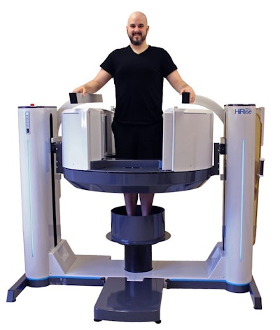
CurveBeam HiRise conebeam CT scanner. Image courtesy of CurveBeam.CurveBeam's HiRise scanner allows clinicians to acquire CT scans displaying the femoral head within the acetabulum in a standing position. When reconstructed into a 3D dataset, the CT scans could facilitate preoperative planning for orthopedic procedures such as knee replacement surgery, the company said.
HiRise has a gantry that elevates and lowers along a vertical track to scan the lower extremities, and tilts 90° to scan the upper extremities. It also comes with an optional table that allows patients to lie down during nonweight-bearing scanning, when necessary.


![Images show the pectoralis muscles of a healthy male individual who never smoked (age, 66 years; height, 178 cm; body mass index [BMI, calculated as weight in kilograms divided by height in meters squared], 28.4; number of cigarette pack-years, 0; forced expiratory volume in 1 second [FEV1], 97.6% predicted; FEV1: forced vital capacity [FVC] ratio, 0.71; pectoralis muscle area [PMA], 59.4 cm2; pectoralis muscle volume [PMV], 764 cm3) and a male individual with a smoking history and chronic obstructive pulmonary disorder (COPD) (age, 66 years; height, 178 cm; BMI, 27.5; number of cigarette pack-years, 43.2, FEV1, 48% predicted; FEV1:FVC, 0.56; PMA, 35 cm2; PMV, 480.8 cm3) from the Canadian Cohort Obstructive Lung Disease (i.e., CanCOLD) study. The CT image is shown in the axial plane. The PMV is automatically extracted using the developed deep learning model and overlayed onto the lungs for visual clarity.](https://img.auntminnie.com/mindful/smg/workspaces/default/uploads/2026/03/genkin.25LqljVF0y.jpg?auto=format%2Ccompress&crop=focalpoint&fit=crop&h=112&q=70&w=112)

