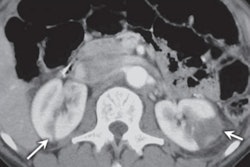
The presence of a reversed halo sign on chest CT scans is indicative of septic pulmonary embolism (PE) in patients with intravenous (IV) substance use disorder, according to an article published online October 31 in the American Journal of Roentgenology.
The group, led by Dr. Renata Almeida from Harvard Medical School, performed a retrospective analysis of chest CT scans acquired at their institution between 2007 and 2017 with a confirmed finding of septic PE due to abuse of IV substances, including opioids. For the 62 cases, nearly 60% had a reversed halo sign -- a focal rounded area of ground-glass opacity surrounded by a ring of consolidation.
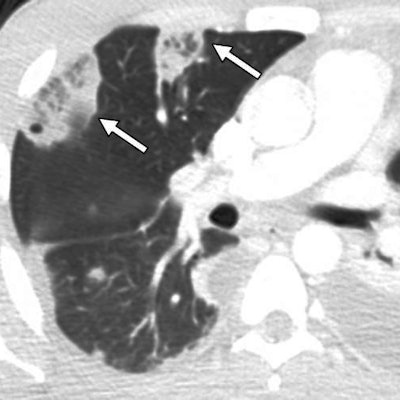
 Chest CT scan showing two reversed halo signs (arrows) with central reticulation in the anterior segment of the right upper lobe. Image courtesy of AJR.
Almeida and colleagues found that the reversed halo sign had a statistically significant association with septic PE in IV drug users (Cohen's kappa coefficient range, 0.84 to 0.96; p < 0.0001). They also found an average of two reversed halo signs per patient.
Reversed halo sign appears to be an "early and reliable imaging finding observed in most cases of CT-based diagnosis of septic PE secondary to IV substance use disorder" and should be included in the differential diagnosis of this patient cohort, the authors concluded.