Monday, December 2 | 3:40 p.m.-3:50 p.m. | SSE14-05 | Room S406B
In this scientific presentation, a team of researchers from Taiwan will report on how an artificial intelligence (AI) algorithm served as a helpful second reader for whole-body CT exams.Nearly 200 whole-body CT scans are performed every day at Chang Gung Memorial Hospital in Taiwan. Of these, approximately 70% are ordered for cancers outside of the lung, but include requests for screening of the lung fields, according to presenter Dr. Gigin Lin, PhD.
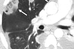
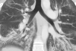
However, interpreting these exams to detect small lung nodules requires extra time for radiologists and hinders them from providing other services in their radiology department, Lin told AuntMinnie.com. In hopes of improving daily workflow and to review the department's false-negative rate for lung nodule detection, the researchers adopted a deep-learning system developed by Ferrum Health to test its diagnostic performance when used as a second reader.
They found that the software could improve workflow and reduce the rate of missed pulmonary nodules. The software found missed pulmonary nodules in 0.3% of the scans; one-third of these misses were deemed to have clinical implications.
The deep-learning system might "have the potential to change treatment decision-making for early detection and early intervention in the era of whole-body CT," Lin said.
Take in this talk on Monday to learn more.