The ambulatory CT unit of Northwestern Medicine Central DuPage Hospital has reduced the amount of time it takes to deliver clot-busting treatment to stroke patients by an average of 30 minutes, according to the health system.
In its first year of service, the mobile stroke unit has enabled clinicians to deliver tissue plasminogen activator (tPA) treatment to ischemic stroke patients in an average of 52 minutes in response to an emergency call, compared with 82 minutes when patients are transported to the hospital for treatment.
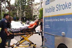
 Northwestern Medicine's mobile stroke unit is equipped with a 16-slice CT scanner. Image courtesy of Northwestern Medicine.
Providing tPA to stroke patients who have a clot is the gold standard, but tPA cannot be offered until the stroke is deemed ischemic and not hemorrhagic on an imaging exam. Equipped with a 16-slice CT scanner, the mobile stroke unit is able to perform on-the-spot imaging and allow clinicians to make that call much earlier than before, according to the health system.
This ambulatory stroke care is available throughout the hospital's emergency medical services area, which includes various suburbs west of Chicago. Northwestern Medicine is working on making this service available to other locations in the vicinity.