A novel radiotherapy technique using conebeam CT (CBCT) shows promise as a treatment for patients with early-stage hepatocellular carcinoma (HCC), according to a study published April 24 in Radiology.
A team led by Dr. Riad Salem of Northwestern University Feinberg School of Medicine found that 90% of patients showed a positive response to the treatment, with 59% of them experiencing a complete response.
Current treatments for the disease include surgery, liver transplantation, and radiofrequency ablation, Salem and colleagues wrote. But many patients don't qualify for these treatments due to other conditions, and they can be costly.
The new technique, called radiation segmentectomy, is a minimally invasive, outpatient procedure that uses the radioisotope yttrium-90 (Y-90) to destroy tumors. Y-90 is embedded into beads that are carried through a catheter into the liver. The beads then deliver radiation to the tumor site while sparing surrounding healthy tissue.
Salem's group studied long-term outcomes in 70 patients with early-stage HCC who had undergone the procedure between 2003 and 2016. Results included the following:
- Nearly 75% of patients had no progression of cancer in the target tumor five years after treatment.
- Median overall survival was 6.7 years.
- One-, three-, and five-year survival probabilities were 98%, 66%, and 57%, respectively.
- In patients with a baseline tumor size of 3 cm or less, one-, three-, and five-year overall survival probabilities were 100%, 82%, and 75%.
"Our results show that we are able to impart curative outcomes to these patients," Salem said in a statement released by the RSNA. "Our numbers with radiation segmentectomy match or outperform those of other curative treatments in terms of tumor control, survival rate, and recurrence."


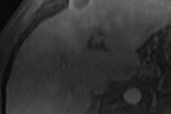

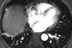


![Axial images from unenhanced calcium score cardiac CT (left) and curved planar reformation images from CT angiography (right) show that higher long-term exposure to air pollution is associated with greater coronary artery calcium and more obstructive coronary artery disease (CAD). Top row: Images in a 68-year-old male patient with higher 10-year mean ambient air pollution exposure (7.9 μg/m3 for particulate matter measuring ≤2.5 μm in diameter [PM2.5] and 17.4 parts per billion [ppb] for NO2) with extensive CAD (coronary artery calcium score [CACS] >1,000 and obstructive CAD [≥70% diameter stenosis]). Bottom row: Images in a 57-year-old female patient with lower 10-year mean ambient air pollution exposure (6.3 μg/m3 for PM2.5 and 4.6 ppb for NO2) with no CAD (CACS = 0 and no obstructive stenosis).](https://img.auntminnie.com/mindful/smg/workspaces/default/uploads/2026/06/hanneman.r6SMLzkezo.png?auto=format%2Ccompress&dpr=2&fit=crop&h=167&q=70&w=250)


