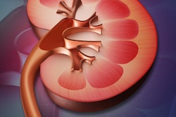
There is no association between IV contrast media used in CT scanning and chronic kidney disease, dialysis, kidney transplants, or acute kidney injury, despite long-held suspicions to the contrary, according to a five-year study published January 25 in the Annals of Emergency Medicine.
Investigators from Johns Hopkins University concluded that since the advent of today's controlled contrast administration regimens, the increased risk of kidney injuries seen in previous studies is simply not present. They believe their research is the largest controlled study to date of acute kidney injury (AKI) following contrast administration (Ann Emerg Med, January 25, 2017).
Physicians have had concerns that contrast media administration can result in serious kidney problems. Indeed, some studies have shown that contrast-induced nephropathy occurs in as many as 14% of patients who get contrast, said lead study author Dr. Jeremiah Hinson, PhD, in a statement accompanying the study's release.
"However, studies used to establish this risk were performed prior to the development of modern contrast reagents or without adequate controls," Hinson said. "Using a controlled design in current context, we could not find an association between intravenous contrast media use and acute kidney injury."
On the other hand, the more than 80 million doses of IV contrast media that are administered every year are essential to accurately diagnosing certain conditions, he added.
The study looked at five years of records for patients who underwent CT exams with or without contrast. Contrast-enhanced exams represented more than half of all studies. The probability of developing acute kidney injury was 6.8% for patients undergoing contrast-enhanced CT, 8.9% for patients receiving unenhanced CT, and 8.1% for patients who were not scanned at all.
A well-controlled, randomized, prospective study is still needed to fully determine the contribution of intravenous contrast media to the development of acute kidney injury, but these results show that in emergency departments with protective measures in place for the use of iodinated contrast, the risk of acute kidney injury is not increased, the study team reported.
The benefits of the timely use of contrast-enhanced CT in avoiding missed diagnosis of critical disease far outweigh any risk of acute kidney injury, the researchers concluded.