Wednesday, December 4 | 11:20 a.m.-11:30 a.m. | SSK05-06 | Room E351
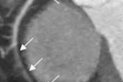
Researchers dialed their CT radiation dose way down -- to 1 mSv per CT colonography exam -- and reconstructed the images using two different iterative reconstruction algorithms in this study from Korea.Their goal was to see if one or both could still visualize the ultralow-dose findings, and to investigate whether computer-aided detection (CAD) could find the polyps.
Using 120 kVp and a photon-starving 10 mAs of tube current, they reconstructed the images using adaptive statistical iterative reconstruction 80% (ASIR, GE Healthcare) and model-based iterative reconstruction (MBIR, GE), running the whole thing through a commercially available CAD scheme.
CAD worked a little better with the second-generation iterative reconstruction algorithm, but not enough to make a significant difference, reported Dr. Eun Sun Lee, PhD.
"Although improvement of CAD sensitivity was slightly bigger with MBIR than with ASIR, we were not able to achieve statistical significance," Lee told AuntMinnie.com. "We thought that a small number of the study population may be responsible for such an insignificant difference, and a further study with a larger study population should be performed to prove the benefit of iterative reconstruction."