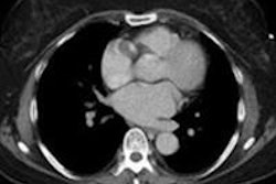
A new article in the September issue of the Journal of the American College of Radiology offers pointers for optimizing radiation dose in chest CT scans, the third most commonly performed CT exam in the U.S.
Dr. Sarabjeet Singh, Dr. Mannudeep Kalra, and Dr. James Thrall, from Massachusetts General Hospital, and Mahadevappa Mahesh, PhD, from Johns Hopkins University, note that optimal dose reduction always starts with ensuring that there is a justifiable clinical indication for performing CT (JACR, September 2011, Vol. 8:9, pp. 663-665).
The lungs are well suited to low-dose imaging in the first place, the authors noted. The low-attenuation background of air-filled lungs offers a high-contrast setting that can be used to assess lung abnormalities at very low radiation dose.
Other recommendations by the authors include the following:
- Patients should receive clear instructions. Technologists should ensure that patients understand breath-hold instructions and avoid voluntary movement during scans.
- Automatic exposure control (AEC) techniques should be used for most chest CT scans in children and adults. Faster gantry rotation times can help minimize breathing artifacts.
- For all but the heaviest patients, localizer radiographs can be acquired at 80 kVp, with the lowest possible tube current of 10 to 20 mA. A pitch of 1.1 or greater improves CT radiation dose efficiency. Lung nodules and most lung-only abnormalities can be evaluated at very low radiation doses compared with mediastinal or other soft-tissue lesions.
- Partial iterative reconstruction techniques, when available, can reduce the necessary radiation dose significantly.
- Centering patients in the gantry isocenter is key to dose optimization; excessive scan length wastes dose. Scan length should be restricted to the area of indication.
- Reading thicker sections will allow for radiation dose reduction while allowing fine details to be read in noisier, thinner sections. Acquiring thin sections and then reconstructing thicker images with multiplanar reformats can also reduce image noise and improve image quality.
- Most standard chest CT scans can be performed at 100 or 120 kVp, with 100 kVp reserved for smaller adult subjects. The American College of Radiology (ACR) recommends diagnostic CT reference volume CT dose indexes of 25 mGy (adult) and 20 mGy (pediatric) abdominal studies, figures that can serve as good reference benchmarks for optimizing standard chest CT protocols.
"The acquisition of images in three phases must not triple radiation dose compared with single-phase chest CT," Singh and colleagues wrote. "Generally, volumetric helical acquisition is preferred for initial inspiratory-phase CT. Subsequently, axial or step-and-shoot mode can be used for expiratory phase and prone images are acquired at 2-cm intervals from the lung apices to the base at low tube current."
The authors also recommended two sources for additional information: www.imagegently.org for low-dose scanning and www.imagewisely.org for radiation safety information.


