Both 64-detector-row CT and 3-tesla MRI are "equally proficient" for pancreatic cancer detection and pancreatic lesion characterization, according to a study in the June issue of Radiology.
Researchers from the Medical University of Vienna found no significant statistical difference between the sensitivity and specificity rankings of two experienced readers interpreting images from both modalities. The authors gave CT a slight advantage over MRI, only because CT is more available in clinical settings.
Lead study author is Dr. Claus Koelblinger from the university's department of radiology.
From September 2006 to November 2007, researchers enrolled 89 patients suspected of having pancreatic cancer who were referred to surgeons at the university based on findings from clinical examination or previous imaging studies. The study included 48 women with a mean age of 65.6 years and 41 men with a mean age of 65.3 years (Radiology, June 2011, Vol. 259:3, pp. 757-766).
CT scans were conducted on a 64-detector-row scanner (Sensation 64, Siemens Healthcare). Patients also received 150 mLof nonionic contrast material (Iomeron 300, Bracco Diagnostics) administered with a power injector.
MRI scans were performed on a 3-tesla system (Trio Tim, Siemens) with a phased-array coil. All patients were injected with a bolus of 0.1 mmol/kg of gadobenate dimeglumine contrast agent (MultiHance, Bracco).
Two gastrointestinal radiologists reviewed the CT and MR images independently and were unaware of all clinical information and final diagnoses. To minimize recall bias, interpretation of images was performed at eight weeks apart and presented in random order.
Focal pancreatic masses were found in 63 patients, with 43 patients diagnosed with adenocarcinoma. Both readers discovered a total of 70 pancreatic lesions, with 56 lesions (80%) determined to be malignant.
For the first reader, sensitivity in the detection of pancreatic adenocarcinoma with CT was 98% (42 of 43 patients), compared with the same 98% for MRI. Specificity with CT and MRI again were identical at 96% (44 of 46 patients).
With the second reader, sensitivity in the detection of pancreatic adenocarcinoma with CT was 93% (40 of 43 patients), compared with 95% for MRI (41 of 43 patients). Specificity for CT and MRI were the same at 96% each (44 of 46 patients).
Diagnostic accuracy for lesion classification on a per-patient basis for the first reader was 94% with both CT and MRI (84 of 89 patients). The second reader had diagnostic accuracy of 91% with CT (81 of 89 patients), compared with 93% with MRI (83 of 89 patients).
Vessel infiltration was determined in 22 patients who underwent surgery, with the first reader achieving sensitivity of 90% with CT (none of 10 vessels), compared with 80% with MRI (eight of 10 vessels). Specificity was 98% (119 of 122 vessels) with CT, compared with 96% with MRI (117 of 122 vessels).
The second reader had a sensitivity of 70% with CT (seven of 10 vessels), compared with 50% with MRI (five of 10 vessels). Specificity was 98% with both CT and MRI (120 of 122 vessels).
 |
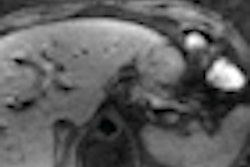
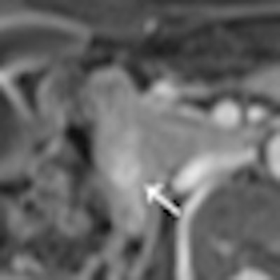
| An ampullary carcinoma of 1.5 mm (arrow) is seen with contrast-enhanced T1-weighted MRI (left), but is not visible on contrast-enhanced CT (right). Image courtesy of Radiology. |
In patients with a normal pancreas, each reader identified one false-positive lesion with CT (specificity of 96%). The first reader found two false-positive lesions with MRI (specificity of 92%), while the second reader discovered one false-positive lesion with MRI (specificity of 96%).
The first reader rated both false-positive MR images as malignant and the false-positive CT finding as benign. The second reader judged the false-positive lesion detected with both modalities as malignant.
Both readers also correctly assessed resectability in 13 of 15 patients (87%) with CT and 14 of 15 cases (93%) with MRI. Nonresectability was assessed correctly with CT in 75% (six of eight patients) of cases by the first readers and 63% (five of eight patients) of cases by the second reader.
By comparison, nonresectability was correctly identified by MRI in 75% (six of eight patients) of cases by the first reader and 50% (four of eight patients) of cases by the second reader. None of the differences between modalities and readers were statistically significant, Koelblinger and colleagues noted.
Based on the results, 64-detector-row CT and 3-tesla MRI are "equally proficient for the detection of pancreatic cancer and pancreatic lesion characterization," they concluded.
The authors, however, cautioned that "some uncertainties still remain with regard to the diagnosis of borderline resectable tumors with close tumor-to-vessel contact and the differentiation between inflammatory pseudotumors and cancer."