VIENNA - Toshiba Medical Systems Europe of Zoetermeer, Netherlands, is highlighting new software applications for functional CT imaging at this week's European Congress of Radiology (ECR).
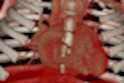
Whole-organ body perfusion makes use of the 16-cm organ coverage of the company's 320-detector-row Aquilion One scanner to enable the visualization of an entire organ in one rotation, adding a time element to organ perfusion studies.
The application enables the acquisition of multiple isophasic images with preset delay times, and users can set the region of interest in portal and/or arterial vessels, according to the company. The software can be useful for clinical applications such as following up on liver transplants.
The second new application, myocardial perfusion, is designed to compensate for difficulties in quantifying cardiac tissue functionality caused by obstructed coronaries or infarcts. Toshiba is again highlighting Aquilion One's 16 cm of coverage as an advantage, as it enables scanning of the entire heart without having to move the patient, resulting in homogeneous contrast distribution.
Toshiba said the application involves comparing rest and stress data. A rest scan is performed using a low-dose, prospective CT angiography protocol. Then, a pharmacological stress agent is administered, and a low-dose stress scan is performed.
A major advantage of the protocol is its ability to complete a cardiac diagnosis with just one exam, according to Toshiba. Commercial deliveries are scheduled to start in August 2010.
Related Reading
Toshiba launches Aquilion cardiac software, March 1, 2010
Toshiba promotes Ryan to VP spot, February 23, 2010
FDA clears Toshiba's Viamo ultrasound, February 4, 2010
Toshiba nets midsized Infinix VF-i install, January 27, 2010
Toshiba brings Aquilion One to London, January 13, 2010
Copyright © 2010 AuntMinnie.com


![Images show the pectoralis muscles of a healthy male individual who never smoked (age, 66 years; height, 178 cm; body mass index [BMI, calculated as weight in kilograms divided by height in meters squared], 28.4; number of cigarette pack-years, 0; forced expiratory volume in 1 second [FEV1], 97.6% predicted; FEV1: forced vital capacity [FVC] ratio, 0.71; pectoralis muscle area [PMA], 59.4 cm2; pectoralis muscle volume [PMV], 764 cm3) and a male individual with a smoking history and chronic obstructive pulmonary disorder (COPD) (age, 66 years; height, 178 cm; BMI, 27.5; number of cigarette pack-years, 43.2, FEV1, 48% predicted; FEV1:FVC, 0.56; PMA, 35 cm2; PMV, 480.8 cm3) from the Canadian Cohort Obstructive Lung Disease (i.e., CanCOLD) study. The CT image is shown in the axial plane. The PMV is automatically extracted using the developed deep learning model and overlayed onto the lungs for visual clarity.](https://img.auntminnie.com/mindful/smg/workspaces/default/uploads/2026/03/genkin.25LqljVF0y.jpg?auto=format%2Ccompress&crop=focalpoint&fit=crop&h=100&q=70&w=100)


![Images show the pectoralis muscles of a healthy male individual who never smoked (age, 66 years; height, 178 cm; body mass index [BMI, calculated as weight in kilograms divided by height in meters squared], 28.4; number of cigarette pack-years, 0; forced expiratory volume in 1 second [FEV1], 97.6% predicted; FEV1: forced vital capacity [FVC] ratio, 0.71; pectoralis muscle area [PMA], 59.4 cm2; pectoralis muscle volume [PMV], 764 cm3) and a male individual with a smoking history and chronic obstructive pulmonary disorder (COPD) (age, 66 years; height, 178 cm; BMI, 27.5; number of cigarette pack-years, 43.2, FEV1, 48% predicted; FEV1:FVC, 0.56; PMA, 35 cm2; PMV, 480.8 cm3) from the Canadian Cohort Obstructive Lung Disease (i.e., CanCOLD) study. The CT image is shown in the axial plane. The PMV is automatically extracted using the developed deep learning model and overlayed onto the lungs for visual clarity.](https://img.auntminnie.com/mindful/smg/workspaces/default/uploads/2026/03/genkin.25LqljVF0y.jpg?auto=format%2Ccompress&crop=focalpoint&fit=crop&h=112&q=70&w=112)


