The ability to depict blood vessels accurately within a bony environment is vital to the correct diagnosis of intracranial vascular pathologies. However, a weak point of conventional CT angiography (CTA) is its limited capacity to depict aneurysms and stenoses located at the skull base, close to the bone.
One possible solution is the use of specialized CT digital subtraction angiography (DSA) software, which enables users to remove bone from images by comparing two separate CT scans, one contrast-enhanced and the other nonenhanced. But the new generation of dual-source CT scanners has created the potential to perform bone removal in a single scan using the system's unique dual-energy imaging capability.
A pair of studies presented at the 2008 European Congress of Radiology (ECR) in Vienna compared the effectiveness of both techniques for bone removal. German researchers found the DSA software to be superior to dual-energy CT, but a report from Japan suggested that the latest advances in dual-energy CTA bone removal may offer greater advantages.
In the first study, Dr. Karsten Papke from the University of Duisburg-Essen in Germany showed that bone removal using the DSA software was "superior in all criteria" when compared with a dual-energy CT technique.
For the first dataset, the group used standard clinical protocols to obtain serial contrast-enhanced and unenhanced head CT scans with a dual-source CT scanner (Somatom Definition, Siemens Healthcare, Erlangen, Germany). Bone removal was performed using Siemens' commercially available syngo Neuro DSA software, which subtracts the unenhanced CT data from the contrast-enhanced CT data.
For the second dataset, the researchers used the same dual-source CT scanner, but they employed the system's dual-energy capability rather than using the DSA software. Because the scanner has two x-ray sources, it is able to collect CT images at two different tube voltages, enabling the distinction of bone from opacified vessels.
The results were scored (from 1 = excellent to 4 = poor) according to image quality and the amount of residual bone after removal, based on maximum intensity projections (MIPs). Artificial narrowing or elimination of arteries within the skull base and of bridging veins located next to the calvarium skull section were detected by direct visual comparison of axial source images obtained before and after bone removal, Papke explained.
Mean image quality for Neuro DSA in all criteria was scored as 1.8, compared with 2.7 for dual-energy CT, indicating a superior performance by DSA. All differences were statistically significant.
Papke noted that a further limitation of dual-energy CT was its tendency to remove more than just the bone -- also parts of the vessels -- which may give false-positive results in CTA. "Small bridging veins tend to be subtracted together with the adjacent bone, implying that occlusion may be present," he said.

With regard to identifying calcified plaques, he added that "in arteries they tend to be overestimated with (dual-energy CT), resulting in the overestimation of stenotic degree or depiction of an occlusion instead of a stenosis."
Based on these findings, "in intracranial CTA, bone subtraction with Neuro DSA should be preferred over bone removal with (dual-energy CT)" as it gave "significantly clearer images," Papke concluded. However, he acknowledged that new techniques and software developments were under way in dual-energy CT and that early indications were "promising."
Results from Japan
Indeed, in a separate study, which used a prerelease version of Siemens' syngo dual-energy bone removal software (due for commercial release this month), researchers in Japan concluded that dual-energy CT bone removal "has the potential to replace bone subtraction CTA" methods based on a biphasic scan.
"Our results show that (dual-energy CT) bone removal was better," said Dr. Yoshiyuki Watanabe of the Kyoto Prefectural University of Medicine. "The advantage of dual-energy CT is that bone removal can be done using one postcontrast scan, whereas the bone subtraction method needs the noncontrast scan to subtract the bone structures from the contrast-enhanced scan, and sometimes a patient's movements between the two scans create difficulties."
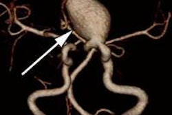
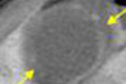
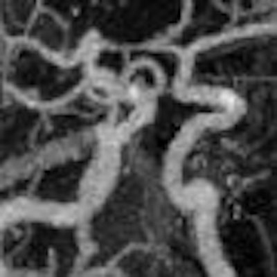
In this study of six patients, three with intracranial aneurysms and three with intracranial carotid stenosis, two aneurysms adjacent to the skull base were only partially visible in conventional CTA, but they were fully visible in dual-energy bone removal CTA. In the three cases of stenosis, conventional CTA failed to display the restricted vessels, while all intracranial stenotic lesions were visible on MIP images from dual-energy CT.
Echoing the findings from Papke's study, however, Watanabe also found that dual-energy CT bone removal led to overestimation of severe stenosis. This was due to hard calcified plaque within stenotic vessels causing x-ray beam hardening, he explained.
"Identifying the borderline between calcification and high iodine concentration was difficult because of the blooming effect," Watanabe said.
The dual-energy CT images were obtained using a Siemens Definition scanner at tube voltages of 80 kVp and 140 kVp, running software that registers the two-energy datasets and selectively removes bone structures. The 3D volume-rendered images and MIP images (with and without bone removal) were reviewed and compared with DSA.
Dual-energy direct bone removal using some of the latest software offers improved diagnostic imaging capabilities over conventional CTA and digital subtraction, Watanabe concluded.
He noted that the technique might also yield benefits in other areas. "Now that ECG-gated dual-energy scanning is available at our institution, the next goal in assessing this (dual-energy) method is to apply the bone removal application for calcified coronary arteries," he said.
By Rob Skelding
AuntMinnie.com contributing writer
April 23, 2008
Related Reading
Patient particulars, subtle clues steer cerebrovascular exam decisions, July 23, 2007
Transient ischemic attacks can be rapidly detected, May 30, 2007
256-slice CT brings new possibilities in head imaging, May 28, 2007
Advanced MR clarifies nexus between white matter and cerebrovascular disease, February 9, 2007
CTA assesses traumatic neurovascular injury, October 16, 2006
Copyright © 2008 AuntMinnie.com