A colorectal cancer screening patient presented with multiple venous malformations that were initially mistaken for neoplastic polyps on virtual colonoscopy, according to a case report published in this month's American Journal of Roentgenology.
"The finding of a polypoid lesion on CT colonography (or virtual colonoscopy) ... has a rather broad differential diagnosis," noted Dr. Andrew Lee, Dr. Perry Pickhardt, and colleagues from the University of Wisconsin Medical School in Madison. "We recently encountered another cause for a polypoid lesion on CT colonography that, to our knowledge, has not been described previously: multiple venous malformations or vascular blebs in an asymptomatic patient with quiescent and previously undiagnosed blue rubber bleb nevus syndrome" (American Journal of Roentgenology, April 2006, Vol. 186:4, pp. 1113-1115).
The report described an asymptomatic 64-year-old man referred for routine VC screening. Following standard cathartic prep consisting of oral sodium phosphate (45 mL), 2% barium sulfate suspension (250 mL), and water-soluble iodinated contrast material (diatrizoate, 60 mL), the patient underwent automated colonic distension with CO2 (ProtoCO2l, E-Z-EM, Lake Success, NY).
Supine and prone CT data were acquired on a 16-detector scanner (LightSpeed, GE Healthcare, Chalfont St. Giles, U.K.), using 1.25-mm collimation, 1-mm reconstruction intervals, 120 kVp, and 50 mAs. The study was read using primary 3D with 2D confirmation on a V3D-Colon workstation (Viatronix, Stony Brook, NY) by a gastrointestinal radiologist with extensive experience in virtual colonoscopy.
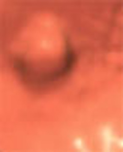
"Multiple polypoid lesions measuring up to 9 mm and located within matching colonic segments were confirmed at optical colonoscopy performed several hours after CT colonography," Andrews et al wrote. "The lesions, however, were not mucosal polyps, but instead were well-defined raised submucosal vascular blebs with a distinctive bluish hue. Biopsy was not performed because of the vascular nature of the lesions, combined with the fact that the endoscopic appearance was characteristic and diagnostic."
 |
| A 64-year-old man was referred for initial colorectal screening. Endoluminal 3D (above and below) and coronal 2D (bottom) images from screening CT colonography showed multiple subcentimeter polypoid lesion (arrows). Images below and bottom show the same lesion on 3D and 2D, respectively. Lesions were all of soft-tissue attenuation and measured up to 9 mm. Images reprinted with permission of the American Roentgen Ray Society (ARRS) from Lee et al, AJR 2006; 186:1113-1115. |
 |
 |
While advanced adenomas are the primary target lesion for colorectal cancer screening, many other entities can present as polypoid lesions at VC, including non-neoplastic mucosal lesions, submucosal lesions, impression from extrinsic lesions, and various VC pitfalls and artifacts.
"Vascular lesions, such as the venous malformations or vascular blebs shown in the current case, represent a rare submucosal cause of polypoid lesions on CT colonography and, to our knowledge, have not been reported previously," Lee et al wrote. "In general, mucosal versus submucosal origin of small focal lesions is sometimes a difficult distinction on CT colonography."
The final diagnosis, blue rubber bleb nevus syndrome, represents a rare condition characterized by multiple venous malformations or hemangiomas involving various organ systems, they wrote.
"Typically, multiple discrete venous lesions manifest primarily in the skin and the gastrointestinal tract, with the small bowel more commonly affected than the large bowel," the group explained. "However, other organ systems may be involved, but not limited to the central nervous, musculoskeletal, genitourinary, and endocrine systems."
After virtual colonoscopy, endoscopy provided the specific diagnosis without need of further workup, they wrote.
By Eric Barnes
AuntMinnie.com staff writer
April 12, 2006
Related Reading
Appendectomy history can lead to false-positive VC, March 1, 2006
Diverticular disease doesn't impede VC in Wisconsin study, November 28, 2005
VC practice thrives in Wisconsin, May 5, 2005
HMO pays for screening virtual colonoscopy, June 4, 2004
Copyright © 2006 AuntMinnie.com


![Axial images from unenhanced calcium score cardiac CT (left) and curved planar reformation images from CT angiography (right) show that higher long-term exposure to air pollution is associated with greater coronary artery calcium and more obstructive coronary artery disease (CAD). Top row: Images in a 68-year-old male patient with higher 10-year mean ambient air pollution exposure (7.9 μg/m3 for particulate matter measuring ≤2.5 μm in diameter [PM2.5] and 17.4 parts per billion [ppb] for NO2) with extensive CAD (coronary artery calcium score [CACS] >1,000 and obstructive CAD [≥70% diameter stenosis]). Bottom row: Images in a 57-year-old female patient with lower 10-year mean ambient air pollution exposure (6.3 μg/m3 for PM2.5 and 4.6 ppb for NO2) with no CAD (CACS = 0 and no obstructive stenosis).](https://img.auntminnie.com/mindful/smg/workspaces/default/uploads/2026/06/hanneman.r6SMLzkezo.png?auto=format%2Ccompress&dpr=2&fit=crop&h=167&q=70&w=250)
