Dynamic x-ray (DXR) imaging can be used to visualize hyperinflation of the lungs in patients with chronic obstructive pulmonary disease (COPD), according to a study published September 29 in the European Journal of Radiology.
The research moves the experimental modality a step forward in clinical development, the authors wrote.
"DXR can generate dynamic sequential images of the thorax with high-temporal resolution," wrote corresponding author Dr. Takuya Hino of Harvard University's Center for Pulmonary Functional Imaging in Boston.
COPD refers to a group of diseases that cause airflow blockage and breathing-related problems, such as emphysema and chronic bronchitis. Spirometry tests to measure airflow limitations are the most common method for diagnosing the disease.
DXR is a technique that has been developed by Konica Minolta that the company is commercializing as dynamic digital radiography (DDR). Previous research showing that the approach can visualize differences in the velocity of airflow in patients with COPD compared with healthy volunteers. The system used in this study was built by Konica Minolta with a flat-panel digital detector (PaxScan 4030CB, Varex Imaging) and a pulsed x-ray generator that transmits a series of x-rays about 15 times per second. Captured static images are then processed by software into dynamic images.
The researchers hypothesized that patients with severe COPD have larger projected lung areas (PLA) on dynamic x-ray imaging compared to healthy people due to hyperinflation of the lungs and that these differences are associated with spirometry metrics used to diagnose the disease, such as tidal volume, normal vital capacity, and forced expiratory volume percent in one second.
The group culled imaging from 32 COPD patients who underwent DXR from June 2009 to August 2011, while 45 healthy volunteers were recruited from May 2013 to February 2014. All participants underwent both spirometry and DXR on the same day. Participants were classified into three groups: normal, COPD mild, and COPD severe.
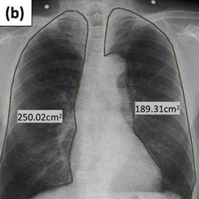
The researchers analyzed associations between spirometry metrics and PLA, which they calculated by manually tracing bilateral lung contours on the DXR images.
Results showed that PLAs were significantly different between the normal and severe COPD groups in both tidal inspiration and expiration and forced inspiration and expiration in both lungs (p ≤ 0.002), according to the findings.
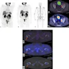
 A 79-year-old male with mild COPD. Projected lung areas (PLA) of (a) tidal inspiratory phase and (b) forced inspiratory phase. Image (a) describes manually located white dots in the left lung field, which were used for automatic calculation of left PLA. Curve graphs describe the temporal change of PLA by automated tracking in (c) tidal and (d) forced respiration. Approximate straight line during forced expiration is colored purple. Image courtesy of the European Journal of Radiology.
PLA in the forced expiratory phase was also different, with a statistical significance between COPD mild and COPD severe groups in the left lung (p < 0.001). In addition, changes in PLA in tidal breathing were lower in normal subjects than in COPD patients (p < 0.001), while changes in PLA in forced breathing were higher in the normal group than in the COPD severe group (p = 0.013), according to the findings.
"COPD severe group has larger projected lung area (PLA) in inspiratory and expiratory phases with tidal and forced respiration in both lungs compared to normal group," the authors wrote.
While several studies have reported the quantification of hyperinflation using chest CT volumetry, no studies have reported the quantification of hyperinflation of lungs with x-ray images, the authors noted.
Ultimately, the DXR technique could eventually enable clinicians to simultaneously assess anatomical and physiological information in patients with COPD, as well as reduce cost and radiation exposure associated with traditional CT imaging, the authors suggest.
Additional research is planned, they wrote.
"Further studies with a large population are required to evaluate the reproducibility of this study," the authors concluded.