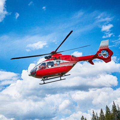
Radiologists are reaching stroke patients by helicopter in remote areas of southeast Bavaria, responding and performing critical procedures in less than an hour in all cases. They discussed the impact of deploying flying intervention teams on patient care in a study published May 5 in JAMA.
In a controlled intervention study, investigators at a telestroke network based in Munich, Germany, deployed flying intervention teams to 15 remote hospitals over a period of about two years beginning in February 2018. The teams performed time-sensitive endovascular thrombectomies (EVTs) on site.
A comparison with time required to transfer patients from remote clinics to the nearest comprehensive stroke unit via ambulance showed the flying teams initiated EVTs in less than half the time (on average, 58 minutes vs. 148 minutes), the authors wrote.
"Because EVT is usually not offered in primary stroke centers, methods to expedite EVT for eligible patients were urgently needed to avoid time-consuming patient interhospital transfer," wrote lead author Dr. Gordian Hubert, a neurologist at the TEMPiS telestroke center in Munich.
EVT for acute ischemic stroke is most effective when applied rapidly after stroke onset to ensure blood supply to affected brain areas, with guidelines recommending initiating treatment as soon as possible. Yet implementation is challenging in rural areas. Lack of neurointerventional expertise is a major barrier in developing EVT-capable hospitals, and long distances to urban comprehensive stroke centers cause critical transfer delays.
A flying intervention team service was established in 2018 for primary stroke centers within the telestroke network of southeast Bavaria (TEMPiS), which covers a 14,000-sq-km area and a population of 2 million. The network treats approximately 5,500 patients with acute stroke per year.
When a patient presents to a primary stroke center and clinical triage reveals the possibility of large-vessel occlusion, an alarm alerts the helicopter crew and flying intervention team to prepare for possible deployment. When the decision to pursue EVT has been made via telemedicine by a neurologist and interventionist, the team and pilots are given a final alarm to deploy.
The intervention team consists of a neurointerventional radiologist and angiography assistant. They bring a special suitcase with intervention-specific items, including rescue material for common complications, and sufficient for three subsequent endovascular procedures. After landing at the local helicopter pad, they are escorted by hospital staff directly to an angiography suite.
Overall, five neurointerventionalists and six assistants participated in the study. The primary outcome was median time from decision to pursue EVT to initiation of procedure (groin puncture) in minutes.
Between February 1, 2018, and October 24, 2019, a total of 157 patients were included in the study, of whom 72 received flying team care and 85 were transferred. EVT was performed in 60 patients (83%) in the flying team group versus 57 (67%) in the transfer group.
The median time from decision to pursue EVT to start of the procedure was 58 minutes (range, 51-71 minutes) in the flying team group and 148 minutes (range, 124-177 minutes) in the transfer group, a difference of 90 minutes, according to the findings.
"Deployment of a flying intervention team to local stroke centers, compared with patient interhospital transfer to referral centers, was significantly associated with shorter time to EVT," the authors wrote.
However, based on follow-up telephone interviews three months after their procedures, the investigators found no statistically significant difference in functional outcomes between the patient groups, the authors noted.
"The findings may support consideration of a flying intervention team for some stroke systems of care, although further research is needed to confirm long-term clinical outcomes and to understand applicability to other geographic settings," Hubert and colleagues concluded.
In an accompanying editorial, Drs. Kori Zachrison and Lee H. Schwamm of Harvard Medical School in Boston wrote that shifting to the use of flying intervention teams will likely lead to improved patient-centered outcomes, including reduced disability and mortality.
"When it comes to stroke, time is brain," they wrote.
Because neuroendovascular specialists are a relatively scarce resource, moreover, bringing the proceduralist to the patient may be an important strategy to maintain high levels of procedural competence in regions with low stroke volumes.
However, achieving robust, standardized, protocol-driven system responses may require different approaches in countries with coordinated, centralized healthcare planning versus countries with more decentralized healthcare delivery, they suggested.
"The imperative to continually strengthen the stroke system of care and increase the value in the delivery of stroke care remains relevant for all, regardless of location," they concluded.