
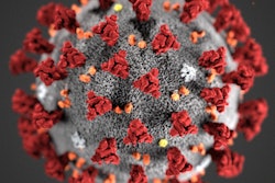
The SARS-CoV-2 virus that causes COVID-19 remains stable for several hours to days when it is aerosolized or on surfaces, according to a letter to the editor published online March 17 in the New England Journal of Medicine.
The virus was found in aerosols for up to three hours, on copper for up to four hours, on cardboard for up to 24 hours, and on plastic and stainless steel for up to two or three days, wrote researchers from the U.S. National Institutes of Health (NIH), the U.S. Centers for Disease Control and Prevention, the University of California at Los Angeles, and Princeton University.
"The results provide key information about the stability of SARS-CoV-2, which causes COVID-19 disease, and suggests that people may acquire the virus through the air and after touching contaminated objects," the team said in an NIH statement.
A group led by Neeltje van Doremalen, PhD, of the U.S. National Institute of Allergy and Infectious Diseases in Hamilton, MT, compared the stability of SARS-CoV-2 with SARS-CoV-1, which causes severe acute respiratory syndrome (SARS) (SARS infected more than 8,000 people around the world in 2002 and 2003 and is the virus most closely related to SARS-CoV-2). The two viruses have similar stability profiles, making it unclear why SARS-CoV-2 appears to be more infectious than its predecessor.
The authors highlighted the following observations:
- It appears that people infected with SARS-CoV-2 might be spreading the virus before they show symptoms, which makes disease control measures that were effective against SARS-CoV-1 less effective now.
- Unlike SARS-CoV-1, most secondary cases of SARS-CoV-2 transmission seem to be occurring in community rather than healthcare settings. But healthcare settings are vulnerable to the introduction and spread of SARS-CoV-2, and its stability in aerosols and on surfaces probably contributes to the transmission of the virus.
"The findings affirm the guidance from public health professionals to use precautions similar to those for influenza and other respiratory viruses to prevent the spread of SARS-CoV-2," the NIH said.





![Examples of ultrasound findings and techniques. (A) Images in a 39-year-old male patient with a mass in the left thigh. The mass is heterogeneous on the B-mode US image (compared with the patient in D) and showed increased microvascularity (superb microvascular imaging [SMI]) and shear-wave elastography (SWE) values. Undifferentiated pleomorphic sarcoma was diagnosed at biopsy (with pleomorphic rhabdomyosarcoma in surgical specimen). (B) Images in an 18-year-old male patient with a mass in the left leg. The mass is hypoechoic on the B-mode image, with no other findings suggestive of malignancy. The lesion is in contact with the cortex of the tibia, which is slightly irregular. CT revealed a doubtful anteromedial tibial erosion. The microvascular study demonstrated high vascularization, suggestive of malignancy. Periosteal Ewing sarcoma was diagnosed with both histologic and immunohistochemical confirmation. (C) Images in a 69-year-old female patient with a lump growing on the outside of the left leg. Multiple SWE examinations were performed (please note the high values obtained in the measurements, whereas the color map highlights the stiffness relative to adjacent tissues). SMI showed areas of increased vascularization to target for sampling. Undifferentiated spindle cell sarcoma was diagnosed at biopsy, with residual leiomyosarcoma in the surgical specimen after neoadjuvant therapy. (D) Images in a 56-year-old female patient with a mass in the right thigh. The mass is heterogeneous at both B-mode ultrasound (similar to patient A) and MRI (coronal T2-weighted spectral attenuated inversion recovery [SPAIR]; T1-weighted pre-contrast and postcontrast imaging), which even shows uptake after the administration of paramagnetic contrast material, which is traditionally suggestive of malignancy. Low values at SMI and elastography are suggestive of benignity. Spindle cell lipoma was diagnosed at biopsy, with atypical spindle cell lipomatous tumor in the surgical specimen.](https://img.auntminnie.com/mindful/smg/workspaces/default/uploads/2026/08/images-radiol250278fig2.APCFLSvX6p.jpg?auto=format%2Ccompress&fit=crop&h=112&q=70&w=112)


