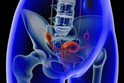
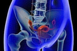
While uterine artery embolization (UAE) has proved an effective interventional radiology treatment for fibroids, concerns remain about reintervention rates years after the initial treatment. Fortunately, a new study has found that UAE reintervention rates are lower than previously thought.
The findings demonstrate that UAE offers women a viable alternative to surgery, wrote a team led by Dr. Ja Kyung Yoon of Yonsei University College of Medicine in Seoul, Korea. The results were published online on October 27 in the European Journal of Radiology
"Uterine artery embolization provides positive mid- and long-term outcomes on symptomatic control and treatment by minimally invasive radiological intervention, and clear advantages over surgery in terms of rapid recovery and return to normal activities," the group wrote.
Uterine fibroids are the most common gynecological tumor, affecting 70% of women by age 50, and they are often treated surgically, according to Yoon and colleagues. Although UAE provides a less-invasive option, its performance over time in terms of further interventions has been a concern: A 2005 study found a reintervention rate of 28.4% at five years and 35% at 10 years.
But it's unclear whether these data are accurate, Yoon's group wrote.
"Although the EMMY trial [A Randomized Comparison of Uterine Artery Embolization and Hysterectomy for the Treatment of Symptomatic Uterine Fibroids] reported outcomes with the longest follow-up, several limitations were noted, such as the higher technical failure rate than present and other reports, lack of information on follow-up MRI, relatively low UAE experience of participating interventional radiologists, and post-UAE management by gynecologists," the researchers wrote. "All of these might have contributed to the reported high intervention rate."
The investigators sought to update the data by conducting a study that investigated the five-year reintervention rate in patients who had undergone UAE. The study included 67 women who underwent bilateral UAE for leiomyoma between March 2011 and February 2012. The women had MRI exams after UAE and again at three-month follow-up. They were then tracked annually, and five years after the procedure, they were surveyed for instances of reintervention.
Fibroid symptoms were resolved in all but one of the 67 women after UAE, for a success rate of 98.5%. Of the women who presented for three-month follow-up and underwent MRI, the fibroid was effectively destroyed in 96.4% of cases. At five-year follow-up, the reintervention rate among the 67 women was 10.4%, with seven cases of further treatment (five myomectomies and two hysterectomies).
Yoon and colleagues also found that a predictive factor for reintervention was the existence of other leiomyomas larger than 2 cm in addition to the primary one that had been treated with UAE.
In any case, the research addresses the concern that UAE's historically high reintervention rates at mid- to long-term follow-up pose a significant limitation to its use, according to the authors.
"This study adds to a body of knowledge that UAE provides an excellent and sustained symptom improvement with a relatively low reintervention rate of 10.4% at five years in the setting of experienced interventional radiology practice," Yoon and colleagues concluded.



![Examples of ultrasound findings and techniques. (A) Images in a 39-year-old male patient with a mass in the left thigh. The mass is heterogeneous on the B-mode US image (compared with the patient in D) and showed increased microvascularity (superb microvascular imaging [SMI]) and shear-wave elastography (SWE) values. Undifferentiated pleomorphic sarcoma was diagnosed at biopsy (with pleomorphic rhabdomyosarcoma in surgical specimen). (B) Images in an 18-year-old male patient with a mass in the left leg. The mass is hypoechoic on the B-mode image, with no other findings suggestive of malignancy. The lesion is in contact with the cortex of the tibia, which is slightly irregular. CT revealed a doubtful anteromedial tibial erosion. The microvascular study demonstrated high vascularization, suggestive of malignancy. Periosteal Ewing sarcoma was diagnosed with both histologic and immunohistochemical confirmation. (C) Images in a 69-year-old female patient with a lump growing on the outside of the left leg. Multiple SWE examinations were performed (please note the high values obtained in the measurements, whereas the color map highlights the stiffness relative to adjacent tissues). SMI showed areas of increased vascularization to target for sampling. Undifferentiated spindle cell sarcoma was diagnosed at biopsy, with residual leiomyosarcoma in the surgical specimen after neoadjuvant therapy. (D) Images in a 56-year-old female patient with a mass in the right thigh. The mass is heterogeneous at both B-mode ultrasound (similar to patient A) and MRI (coronal T2-weighted spectral attenuated inversion recovery [SPAIR]; T1-weighted pre-contrast and postcontrast imaging), which even shows uptake after the administration of paramagnetic contrast material, which is traditionally suggestive of malignancy. Low values at SMI and elastography are suggestive of benignity. Spindle cell lipoma was diagnosed at biopsy, with atypical spindle cell lipomatous tumor in the surgical specimen.](https://img.auntminnie.com/mindful/smg/workspaces/default/uploads/2026/08/images-radiol250278fig2.APCFLSvX6p.jpg?auto=format%2Ccompress&fit=crop&h=100&q=70&w=100)


![Examples of ultrasound findings and techniques. (A) Images in a 39-year-old male patient with a mass in the left thigh. The mass is heterogeneous on the B-mode US image (compared with the patient in D) and showed increased microvascularity (superb microvascular imaging [SMI]) and shear-wave elastography (SWE) values. Undifferentiated pleomorphic sarcoma was diagnosed at biopsy (with pleomorphic rhabdomyosarcoma in surgical specimen). (B) Images in an 18-year-old male patient with a mass in the left leg. The mass is hypoechoic on the B-mode image, with no other findings suggestive of malignancy. The lesion is in contact with the cortex of the tibia, which is slightly irregular. CT revealed a doubtful anteromedial tibial erosion. The microvascular study demonstrated high vascularization, suggestive of malignancy. Periosteal Ewing sarcoma was diagnosed with both histologic and immunohistochemical confirmation. (C) Images in a 69-year-old female patient with a lump growing on the outside of the left leg. Multiple SWE examinations were performed (please note the high values obtained in the measurements, whereas the color map highlights the stiffness relative to adjacent tissues). SMI showed areas of increased vascularization to target for sampling. Undifferentiated spindle cell sarcoma was diagnosed at biopsy, with residual leiomyosarcoma in the surgical specimen after neoadjuvant therapy. (D) Images in a 56-year-old female patient with a mass in the right thigh. The mass is heterogeneous at both B-mode ultrasound (similar to patient A) and MRI (coronal T2-weighted spectral attenuated inversion recovery [SPAIR]; T1-weighted pre-contrast and postcontrast imaging), which even shows uptake after the administration of paramagnetic contrast material, which is traditionally suggestive of malignancy. Low values at SMI and elastography are suggestive of benignity. Spindle cell lipoma was diagnosed at biopsy, with atypical spindle cell lipomatous tumor in the surgical specimen.](https://img.auntminnie.com/mindful/smg/workspaces/default/uploads/2026/08/images-radiol250278fig2.APCFLSvX6p.jpg?auto=format%2Ccompress&fit=crop&h=112&q=70&w=112)