NASHVILLE – Faced with declining membership, revenues, and services, outgoing Association of Educators in Radiological Sciences (AERS) chairperson Donna Lee Wright proposed yesterday that the organization adopt an advocacy-based strategic planning model.
Wright, a research associate professor at the University of North Carolina in Chapel Hill, challenged AERS members at this week's American Healthcare Radiology Administrators (AHRA) conference to embrace "a process by which we voluntarily and continually adjust our organizational strengths to meet the changing needs of our present and future members."
Wright endorsed the advocacy-based planning model based on a presentation made by Howard W. Schwartz at the AERS board of directors mid-year board meeting in January. As a result of the meeting, Wright and her fellow board members took a hard look at their strategic planning policy.
"Traditional strategic planning is based on SWOT: strengths, weaknesses, opportunities, and threats. However, SWOT doesn’t provide much flexibility, it doesn’t allow for rapid response to change. And with so much changing so fast in our field, it’s a planning process that simply doesn’t work," Wright said.
The advocacy model identifies "core values" that will be provided to members in the form of services, products, and benefits. It assigns an individual advocate to protect each core value and act as its champion. The effect will be to evolve the AERS board of directors from being individuals elected as officers to being a collection of advocates for core values identified by members. In addition, the group's size will likely be smaller -- and members will serve longer terms -- than on the current board.
For example, rather than electing a treasurer, members will elect a financial resources advocate. This advocate would oversee budgets and accounts and would also coordinate resource generation.
Wright, along with fellow presenters Nadia Bugg and Rick Carlton, offered three strategic-planning questions for attendees to review with their respective organizations and institutions:
- Why are your current customers still customers?
- What do you need to continue to provide to your customers?
- What new products and services should you provide to your customers?
AERS members also got some good news. Wright reported that expenses are at a minimum, that many members want to make the advocacy-based model work, and that the plan will allow AERS members to set their own priorities for the organization.
A step-by-step transition plan was announced to help ease the organization into the new model. Reorganization efforts will culminate with the election of the first advocates at the 2002 AERS meeting.
By Jonathan S. BatchelorAuntMinnie.com staff writer
August 8, 2000
Let AuntMinnie.com know what you think about this story.
Copyright © 2000 AuntMinnie.com


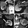
![Examples of ultrasound findings and techniques. (A) Images in a 39-year-old male patient with a mass in the left thigh. The mass is heterogeneous on the B-mode US image (compared with the patient in D) and showed increased microvascularity (superb microvascular imaging [SMI]) and shear-wave elastography (SWE) values. Undifferentiated pleomorphic sarcoma was diagnosed at biopsy (with pleomorphic rhabdomyosarcoma in surgical specimen). (B) Images in an 18-year-old male patient with a mass in the left leg. The mass is hypoechoic on the B-mode image, with no other findings suggestive of malignancy. The lesion is in contact with the cortex of the tibia, which is slightly irregular. CT revealed a doubtful anteromedial tibial erosion. The microvascular study demonstrated high vascularization, suggestive of malignancy. Periosteal Ewing sarcoma was diagnosed with both histologic and immunohistochemical confirmation. (C) Images in a 69-year-old female patient with a lump growing on the outside of the left leg. Multiple SWE examinations were performed (please note the high values obtained in the measurements, whereas the color map highlights the stiffness relative to adjacent tissues). SMI showed areas of increased vascularization to target for sampling. Undifferentiated spindle cell sarcoma was diagnosed at biopsy, with residual leiomyosarcoma in the surgical specimen after neoadjuvant therapy. (D) Images in a 56-year-old female patient with a mass in the right thigh. The mass is heterogeneous at both B-mode ultrasound (similar to patient A) and MRI (coronal T2-weighted spectral attenuated inversion recovery [SPAIR]; T1-weighted pre-contrast and postcontrast imaging), which even shows uptake after the administration of paramagnetic contrast material, which is traditionally suggestive of malignancy. Low values at SMI and elastography are suggestive of benignity. Spindle cell lipoma was diagnosed at biopsy, with atypical spindle cell lipomatous tumor in the surgical specimen.](https://img.auntminnie.com/mindful/smg/workspaces/default/uploads/2026/08/images-radiol250278fig2.APCFLSvX6p.jpg?auto=format%2Ccompress&fit=crop&h=100&q=70&w=100)


![Examples of ultrasound findings and techniques. (A) Images in a 39-year-old male patient with a mass in the left thigh. The mass is heterogeneous on the B-mode US image (compared with the patient in D) and showed increased microvascularity (superb microvascular imaging [SMI]) and shear-wave elastography (SWE) values. Undifferentiated pleomorphic sarcoma was diagnosed at biopsy (with pleomorphic rhabdomyosarcoma in surgical specimen). (B) Images in an 18-year-old male patient with a mass in the left leg. The mass is hypoechoic on the B-mode image, with no other findings suggestive of malignancy. The lesion is in contact with the cortex of the tibia, which is slightly irregular. CT revealed a doubtful anteromedial tibial erosion. The microvascular study demonstrated high vascularization, suggestive of malignancy. Periosteal Ewing sarcoma was diagnosed with both histologic and immunohistochemical confirmation. (C) Images in a 69-year-old female patient with a lump growing on the outside of the left leg. Multiple SWE examinations were performed (please note the high values obtained in the measurements, whereas the color map highlights the stiffness relative to adjacent tissues). SMI showed areas of increased vascularization to target for sampling. Undifferentiated spindle cell sarcoma was diagnosed at biopsy, with residual leiomyosarcoma in the surgical specimen after neoadjuvant therapy. (D) Images in a 56-year-old female patient with a mass in the right thigh. The mass is heterogeneous at both B-mode ultrasound (similar to patient A) and MRI (coronal T2-weighted spectral attenuated inversion recovery [SPAIR]; T1-weighted pre-contrast and postcontrast imaging), which even shows uptake after the administration of paramagnetic contrast material, which is traditionally suggestive of malignancy. Low values at SMI and elastography are suggestive of benignity. Spindle cell lipoma was diagnosed at biopsy, with atypical spindle cell lipomatous tumor in the surgical specimen.](https://img.auntminnie.com/mindful/smg/workspaces/default/uploads/2026/08/images-radiol250278fig2.APCFLSvX6p.jpg?auto=format%2Ccompress&fit=crop&h=112&q=70&w=112)