Researchers from the Massachusetts Institute of Technology (MIT) have developed a wearable breast ultrasound device that they say can make way for earlier breast cancer detection.
The device can image as deep as 15 cm into breast tissue and can image the entire breast from two or three locations, according to research published by the MIT team in Advanced Healthcare Materials. The team described the technology as an end-to-end system ultrasound architecture consisting of a novel sparse array geometry and a codesigned data acquisition system.
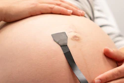
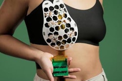
 A new imaging system developed by MIT researchers consists of a small ultrasound probe (left) attached to an acquisition and processing module that is a little larger than a smartphone.Conformable Decoders Lab at the MIT Media Lab
“Our architectural approach enables high performance, real-time 3D ultrasound, removing a key and persistent blocker for the next generation of medical devices,” wrote a team led by Canan Dağdeviren, PhD, and co-authors.
Wearable breast ultrasound devices continue to be explored as a novel method for finding interval cancers early. Dağdeviren and colleagues in 2023 developed an array of ultrasound transducers that were incorporated into a flexible patch that can be attached to a bra. People who wear the device could move an ultrasound tracker along the patch and image the breast tissue from different angles.
However, they noted the potential for small gaps in image coverage that could miss small abnormalities and the need for a connection with traditional image processing machines.
For their new study, the researchers developed a modified ultrasound array that is fully portable and could create a 3D image of the entire breast by scanning two or three locations.
The system is a chirped data acquisition system (cDAQ) that consists of an ultrasound probe and a motherboard that processes the data. It also employs convolutional optimally distributed array (CODA) geometry that reduces the number of elements from 1,024 to 128.
The probe is slightly smaller than a deck of cards and houses an ultrasound array arranged in the shape of an empty square. This allows the array to take 3D images of the tissue below it. The motherboard, meanwhile, processes the acquired data. The motherboard is roughly the size of a smartphone and costs about $300, MIT said. It can be connected to a laptop for viewing images.
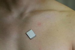
 The probe, which is a little smaller than a deck of cards, contains an ultrasound array arranged in the shape of an empty square, a configuration that allows the array to take 3D images of the tissue below.Conformable Decoders Lab at the MIT Media Lab
The university said the device can be powered with a 5V DC supply, which is used for small electronics.
The researchers tested the new system on a 71-year-old woman with a history of breast cysts. They reported success with the system, highlighting its accuracy in imaging the cysts and creating a 3D image of the breast tissue. The researchers also reported no gaps in image acquisition.
The team also highlighted the device’s placement on top of the skin without having to be pressed into the tissue like a typical ultrasound probe. This means the images are not distorted when they are acquired.
The study authors said this device could help address operator dependence in ultrasound imaging.
“Integrating CODA-based 3D systems with intelligent guidance and automation could help overcome this skill barrier and expand access to advanced diagnostic imaging,” they wrote.
The researchers are conducting a larger clinical trial to further test the device’s capabilities and applications.
Read the full study here.

![A normal mammogram confirmed by three-year radiologic follow-up illustrates reader-marked regions of interest (ROIs) during (A) unaided (round 1) and (B) artificial intelligence (AI)–assisted (round 2) reading. Each colored dot represents an ROI for recall by a human reader. Readers could mark more than one ROI per case, represented by multiple dots of the same color. During AI-assisted reading, the AI system displayed three visible prompts: two with suspicion of malignancy scores of 35% (left mediolateral oblique [L MLO] and craniocaudal [L CC]) and one with a suspicion of malignancy score of 10% (right craniocaudal [R CC]), shown as polygonal overlays. Without AI, six of 10 readers (60%) marked a false-positive ROI. With AI assistance, this fell to two of 10 (20%). R MLO = right mediolateral oblique.](https://img.auntminnie.com/mindful/smg/workspaces/default/uploads/2026/07/2026-07-14-radiology-mammogram-ai-auto-bias.H0bYO8QlWs.jpg?auto=format%2Ccompress&fit=crop&h=100&q=70&w=100)


![A normal mammogram confirmed by three-year radiologic follow-up illustrates reader-marked regions of interest (ROIs) during (A) unaided (round 1) and (B) artificial intelligence (AI)–assisted (round 2) reading. Each colored dot represents an ROI for recall by a human reader. Readers could mark more than one ROI per case, represented by multiple dots of the same color. During AI-assisted reading, the AI system displayed three visible prompts: two with suspicion of malignancy scores of 35% (left mediolateral oblique [L MLO] and craniocaudal [L CC]) and one with a suspicion of malignancy score of 10% (right craniocaudal [R CC]), shown as polygonal overlays. Without AI, six of 10 readers (60%) marked a false-positive ROI. With AI assistance, this fell to two of 10 (20%). R MLO = right mediolateral oblique.](https://img.auntminnie.com/mindful/smg/workspaces/default/uploads/2026/07/2026-07-14-radiology-mammogram-ai-auto-bias.H0bYO8QlWs.jpg?auto=format%2Ccompress&dpr=2&fit=crop&h=167&q=70&w=250)