Minnies finalists, page 3
Scientific Paper of the Year
 Standalone AI for breast cancer detection at screening digital mammography and digital breast tomosynthesis: A systematic review and meta-analysis. Yoon, JH et al, Radiology, May 23, 2023. To learn more about this paper, click here.
The first study the Minnies expert panel picked for Scientific Paper of the Year revealed a fine line – AI algorithms are indeed valuable tools, but their performance raises the uncomfortable specter of radiologists losing their jobs.
In a study published in Radiology, a team led by Dr. Jung Hyun Yoon, PhD, from Yonsei University in Seoul, conducted a meta-analysis including over 1.1 million screening breast imaging exams that used AI software to interpret imaging. AI had a higher pooled area under the curve (AUC) than radiologists (0.87 vs. 0.81) when it comes to mammograms and DBT exams, they found.
In an accompanying editorial, Anabel Scaranelo, MD, of the University of Toronto, urged caution. She wrote that breast imaging departments will need to balance the promise of such tools for reducing fatigue and burnout with "the threat of radiologists losing their jobs to powerful algorithms."
Diagnostic imaging utilization in the emergency department: Recent trends in volume and radiology work relative value units. Poyiadji N et al, Journal of the American College of Radiology, August 3, 2023. To learn more about this paper, click here.
 The second finalist for Scientific Paper of the Year took a deep dive into the increasing demand for advanced imaging in emergency departments. The researchers asked, "What effect is this having on radiologist work relative value units (wRVUs)?"
The answer isn't great, considering the current radiology workforce shortage, they noted.
A team led by Neo Poyiadji, MD, from Henry Ford Hospital in Detroit, analyzed emergency department imaging use volumes and wRVUs between 2014 and 2021. The results illustrated increases of 35.5% in CT volumes at their level one trauma center and a 53.7% spike in wRVUs; meanwhile, MRI volumes increased by 56.3% and wRVUs by 69.4%.
The authors speculated that many factors could play a part in these trends, including fear of malpractice litigation and the desire to bypass preauthorization requirements for advanced imaging.
Perhaps most importantly, however, they suggested that this trend will likely continue.
Best New Radiology Device
BodyTom 64 point-of-care mobile CT scanner, NeuroLogica 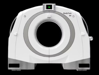 NeuroLogica's BodyTom 64 whole-body, portable CT scanner. Image courtesy of NeuroLogica.
NeuroLogica unveiled its BodyTom 64 point-of-care mobile CT scanner in November 2022 after receipt of U.S. Food and Drug Administration 510(k) clearance.
An upgraded version of the company's BodyTom Elite CT scanner, BodyTom 64 is a 64-slice, full-body portable scanner that’s suitable for use in multiple departments and for both pediatric and adult imaging. For example, it can optimize interventional radiology workflows by remaining ready to rescan for each stage of needle guidance, according to the Samsung Electronics subsidiary.
It can also make use of a radiolucent skull fixation device to enable intraoperative neuroimaging. Thanks to its internal lead shielding and battery operation, it can also be deployed in trauma bays, NeuroLogica said. The firm has also incorporated a number of changes to the software that had been utilized in BodyTom Elite, including switching to a Linux operating system.
Signa PET/MR AIR scanner, GE HealthCare
 GE HealthCare's Signa PET/MR AIR scanner. Image courtesy of GE HealthCare.GE HealthCare introduced its Signa PET/MR AIR scanner at the annual Society of Nuclear Medicine and Molecular Imaging (SNMMI) conference in June.
Signa PET/MR AIR features the vendor’s Adaptive Image Receive (AIR) technologies, including AIR Coils for improved patient comfort; AIR Recon DL for deep learning-based MR image quality improvement and up to 50% faster scan times; and MotionFree Brain software for mitigating motion-related PET image degradation, according to the vendor.
GE HealthCare has also pointed to the system’s time-of-flight capabilities, which the company believes can assist clinicians in delivering early diagnosis, assessing disease progression, and detecting adverse effects in patients with Alzheimer’s disease. What’s more, Signa PET/MR AIR offers particular value in detecting and staging/restaging of prostate cancer, as well as helping to select patients for subsequent prostate-specific membrane antigen (PSMA)-targeted radioligand therapy, according to the firm.
Best New Radiology Software
AWS HealthImaging platform, Amazon Web Services
Amazon Web Services (AWS) made a splash this summer with the launch of its AWS HealthImaging cloud-based imaging data platform.
Designed to reduce the amount of infrastructure required to operate enterprise imaging systems. AWS HealthImaging supports storage, access, and analysis of medical images at a petabyte scale, according to the vendor.
 AWS HealthImaging being used to store an image within an open-source viewer. Image courtesy of Amazon Web Services.
After taking in DICOM data, the platform provides application programming interfaces for low-latency retrieval and purpose-built storage, AWS said. All of an organization’s applications can access a single copy of data without duplication. AWS believes its platform could potentially decrease the total cost of medical imaging storage by up to 40%.
Omni Reporting, Rad AI
Announced in August, Rad AI’s Omni Reporting AI-based radiology reporting software utilizes generative AI technology to help save radiologists time in dictating reports.
Omni Reporting, which is designed to replace existing reporting software products, also helps radiologists improve their report accuracy and consistency, Rad AI said. What’s more, the company has also included its Omni Impressions software for automated report impressions.
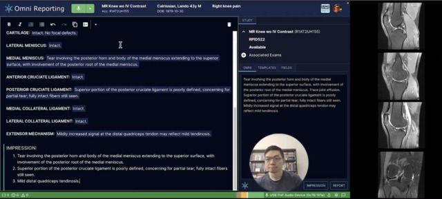 Rad AI's Omni Reporting software used generative AI technology to assist radiologists in preparing radiology reports. Image courtesy of Rad AI.
Omni Reporting is scheduled to be released commercially at this year’s RSNA meeting in Chicago.
Best New Radiology Vendor
Coreline Soft
This South Korean AI and advanced visualization software developer has developed a range of software applications focused largely on lung imaging.
Coreline’ Soft's Aview product line features its LCS lung cancer screening software, which received U.S. Food and Drug Administration (FDA) 510(k) clearance in April and Health Canada approval in June. Designed to help clinicians identify and characterize lung nodules on low-dose CT exams, LCS has been shown to decrease imaging interpretation time by 70%, increase sensitivity by 34%, and lower false positives by 42%, according to the vendor.
 Coreline Soft's LCS Plus software for analysis of CT lung cancer screening exams. Image courtesy of Coreline.
And in July, the company secured FDA clearance for its RT ACS radiotherapy autocontouring software. In other recent developments, research presented at the RSNA 2022 meeting showed that Coreline’s Aview software could improve CT assessment of patients with idiopathic pulmonary fibrosis.
Other Aview software applications developed by Coreline include: CAC deep learning-based software for automatic scoring of coronary artery calcium (CAC) on low-dose CT exams; COPD software for analysis of chronic obstructive pulmonary disease (COPD) phenotypes; Lung Texture software for automated analysis of interstitial lung disease; Interstitial Lung Abnormalities (ILA) analysis software, and NeuroCAD software for automatic detection of cerebral hemorrhage on brain CT.
LifeVoxel
LifeVoxel has developed AImagine, an AI and advanced visualization software platform.
Available as a software-as-a-service, AImagine utilizes predictive techniques and optimized graphical processing methods to provide medical professionals with fast access to diagnostic-quality images, according to the vendor.
In other features, AImagine uses AI algorithms to predict how users will move or manipulate images; rendered images are then transmitted to the user’s device before they have even been requested, according to the vendor. LifeVoxel said it operates three dedicated graphics processing unit (GPU) server farms to provide hosting services for AImagine.
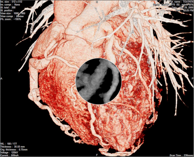 LifeVoxel's software can provide adaptive visualization of the heart using AI for direct physician use, according to the company, Shown here is the left anterior view (LAO) of the heart with focus on left anterior descending artery (LAD) with magnified multiplanar reformatting (MPR) view demonstrating focal narrowing of the LAD proximal to mid-section. Image courtesy of LifeVoxel.
After receiving $5 million in seed funding in 2022, LifeVoxel launched its Proctor telepresence surgery package later that year. In 2023, LifeVoxel rolled out its Providence diagnostic app marketplace and has also unveiled its Telemergency holographic AI technology.
Kovey Kovalan serves as CEO and founder.
Best Educational Mobile App
CTisus Chest Atlas 3D CRT, Elliot Fishman, MD, (iOS)
Although it’s the first time CTisus Chest Atlas 3D CRT has achieved finalist status, other CTisus apps developed by Elliot Fishman, MD, of Johns Hopkins University have been regular contenders for Best Educational Mobile App over the years.
CTisus iQuiz went on to win in 2020, while CTisus iPearls was awarded the Minnie in 2019 and 2017. CTisus Critical Diagnostic Measurements in CT was the first CTisus winner in 2017.
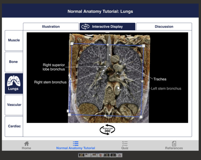 Normal chest anatomy tutorial on CTisus Chest Atlas 3D CRT. Image courtesy of Elliot Fishman, MD.
CTisus Chest Atlas 3D CRT is designed to reveal the unique advantages of cinematic rendering technique (CRT), an imaging method that creates photorealistic 3D images. Focusing on chest anatomy, the app includes a tutorial that establishes a pattern-based approach and includes 360-degree visualization followed by a self-assessment section.
Launched in 2020, CTisus Chest Atlas has recently been upgraded to version 3.0, which includes an updated user interface.
Radiology Assistant 2.0, BestApps (iOS)
A six-time Minnies finalist and winner in 2018 and 2021, Radiology Assistant 2.0 has once again made it to the finals.
Developed by Dutch radiologists Wouter Veldhuis, MD, PhD, of the University Medical Center Utrecht and Robin Smithuis, MD, of Rijnland Hospital Leiderdorp, Radiology Assistant 2.0 features peer-reviewed articles written by expert radiologists. Smithuis is also the founder of the Radiology Assistant educational website and nonprofit organization.
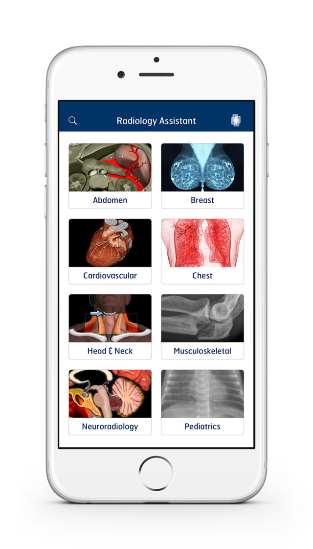 Radiology Assistant 2.0 features articles written by expert radiologists on a variety of categories. Image courtesy of BestApps.
Radiology Assistant 2.0 was first released in 2017 and has been regularly updated over the years. Improved DarkMode support was added in the app's latest update.
Best Radiology Image
The first finalist for Best Radiology Image is from research showing that photon-counting CT (PCCT) effectively detects heart disease in high-risk patients. In a study published June 20 in Radiology, lead author Muhammad Hagar, MD, from the University of Freiburg in Germany described how the modality offers "significant benefit for people previously ineligible for noninvasive screening” and could help clinicians better diagnose heart disease and tailor patient care.
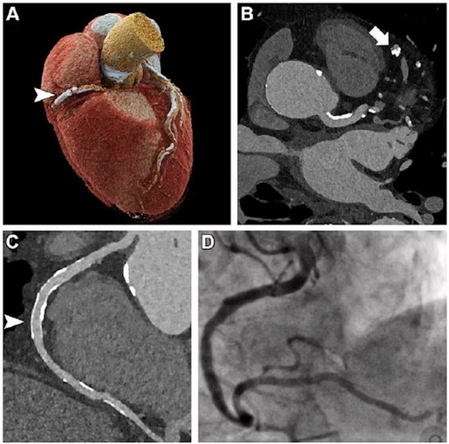 Ultrahigh-resolution (UHR) coronary CT angiography (CCTA) in an 85-year-old man before transcatheter aortic valve replacement. Despite a stent in the right coronary artery and very severe coronary sclerosis with an Agatston score of 4,162, diagnostic visualization of the coronary arteries succeeded, and obstructive coronary artery disease was excluded on CT images. (A) Three-dimensional cinematic rendering of the heart. The stent (arrowhead) is visible in the middle segment of the right coronary artery. (B) UHR CCTA with 0.2-mm axial sections. The lumen (arrow) of the severely calcified distal left anterior descending artery can be assessed without artifacts. (C) Curved multiplanar reformations of the right coronary artery with a diagnostic display of the stent lumen (arrowhead). (D) Invasive coronary angiography enables exclusion of in-stent stenosis. Image and caption courtesy of the RSNA.
The second finalist for Best Radiology Image comes from Stanford University and shows the significant advantages of both PET and SPECT for prostate cancer patients, and how much faster SPECT acquisitions will complement other developments in the field of theranostics, according to Andrei Iagaru, MD, chief of nuclear medicine and molecular imaging told AuntMinnie.com.
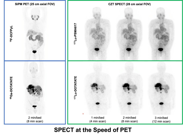 Top row shows maximum intensity projection (MIP) images acquired after injection of diagnostic F-18 DCFPyL using PET and therapeutic lutetium-177 (Lu-177)-PSMA-617 using SPECT in a patient with prostate cancer. Bottom row shows MIP images acquired after injection of diagnostic gallium-68 (Ga-68) DOTATATE using PET and therapeutic Lu-177-DOTATATE using SPECT in a patient with a neuroendocrine tumor. Image montage and caption information courtesy of Andrei Iagaru, MD.
“While fast PET (1-2 minutes per bed for an average of 8-10 minutes scan) has been possible since the introduction of PET/CT scanners with detectors using silicon photomultipliers, it is the recent introduction of SPECT/CT scanners with detectors using cadmium-zinc-teluride (CZT) positioned around the patients that allow for very fast image acquisitions after treatments using beta emitters such as [Lu-177],” Iagaru told AuntMinnie.com. “Scans acquired as fast as 4 minutes with image quality comparable to those acquired in 8 or 12 minutes will improve patient convenience and allow for documentation of treatment distribution and personalized dosimetry, particularly in clinics with high patient volumes.”
Continue reading: Back to Page 1, Back to Page 2.