CEA-Scan Positive 5 Months Before CT
This is a 77 year-old male who presented with rectal cancer in 1995. After surgery, the patient underwent 5 months of chemotherapy and a regimen of radiation therapy as well. Unfortunately, however, in March of 1997, local recurrence was detected and he underwent a second surgery. One year later, the blood CEA level rose to 7 ng/ml. A CT scan was performed, revealing an "ill-defined presacral density and perirectal thickening possibly due to scarring, surgery, or recurrent neoplasm". A follow-up CT was scheduled for 3 months to consider if further evaluation was warranted.
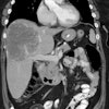
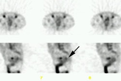
In June of 1998, the next CT showed the density was unchanged since the prior CT scan. The blood CEA level began to rise during the following spring (March 1999) and a CT with a CEA-Scan were ordered. CT revealed a "stable density in the presacral area, most likely representing scarring", however CEA-Scan SPECT images showed a "large focal area of increased uptake in the rectal area with a central photopenic area highly suggestive of rectal recurrence". Since the uptake of CEA-Scan appears to correspond with the mass seen on CT a CT-guided needle biopsy was ordered, however, the biopsy results were negative for malignancy. Another CT was made in September of 1999 that revealed a gross enlargement of the pre-sacral mass, suggestive of recurrence.
Discussion:
It is not uncommon for CEA-Scan to have positive uptake before the CT
has noticeable anatomic changes. This is due to the fact that an anatomic scan like CT is
dependent upon tissue displacement, while CEA-Scan localizes in those areas that express
CEA. This case illustrates how Nuclear Imaging studies and CEA-Scan can often detect
physiologic changes before anatomic changes take place.


Performed on an ADAC camera, this case is from St. Joseph's Hospital in Nashua, NH. The radiologist is Dr. Nicholas Hoff.