
A group in Ireland has used data from dual-energy x-ray absorptiometry (DEXA) scans to estimate the country's prevalence of osteoporosis, with findings suggesting an urgent need to establish a national program to reduce the illness burden.
Researchers led by John Carey, MD, professor in rheumatology at the National University of Ireland and clinical lead in DXA, osteoporosis, and fracture liaison services at Galway University Hospitals, estimated that in 2022, more than 1 million Irish adults over 50 years old have low bone mass and more than 250,000 have osteoporosis.
"We have shown for the first time in a large Irish population that low bone mass defined by [DEXA] classification is common in men and women aged ≥ 50 years, whether they have risk factors or not, or previous osteoporotic fractures," the group wrote, in an article published August 10 in JBMR Plus.
Ireland has one of the largest proportional illness burdens related to osteoporosis in Europe, and it is projected to have the greatest increase over the coming decade, the researchers wrote. Moreover, the cost of managing those who fracture will double by 2030.
DEXA-measured bone mineral density (BMD) is an established criteria to help understand the epidemiology of osteoporosis and identify those at risk for fracture. In this study, the researchers estimated prevalence in Ireland based on DEXA metrics identified in men and women who had scans between January 2000 and November 2018 as part of an ongoing epidemiological study.
A total of 33,344 subjects met the study's inclusion criteria. Among the cohort, the group compared the prevalence of DEXA low bone mass (T-score < -1.0) and of osteoporosis (T-score ≤ -2.5) in adults without major risk factors or fractures, with one or more major risk factors, and with one or more major osteoporotic fractures.
 A graphical abstract. Image courtesy of JBMR Plus.
According to the findings, more than one-half of the men and women without fractures had low bone mass (T-score < -1.0) at one or more skeletal sites, whereas almost one in five had osteoporosis (T-score ≤ -2.5) at one or more skeletal sites. The majority of those with a previous major osteoporotic fracture had low bone mass, whereas only one in three had a T-score less than or equal to -2.5.
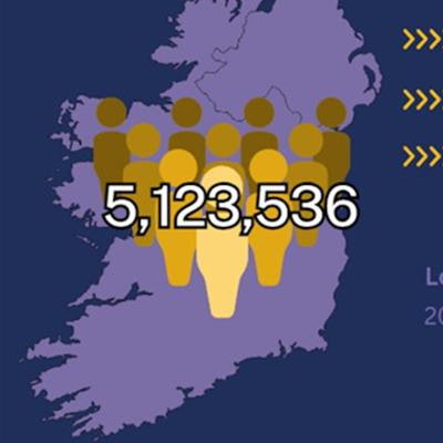
Applying these results to Ireland's 2022 census figure of approximately 5.1 million people, the researchers estimated that between 1 million men and 1.2 million women over 50 years old have low bone mass, whereas between 308,474 men and 498,104 women have osteoporosis.
"We describe for the first time a reasonable estimate of the true prevalence of low bone mass and osteoporosis in a large cohort of older Irish adults using Irish data," the group wrote.
Ultimately, the study highlights that DEXA-measured BMD remains an appropriate choice for clinicians to diagnose osteoporosis in perimenopausal or postmenopausal women, or men ages 50 and older without a major osteoporotic fracture, the researchers noted. Future projects will assess the validity of various fracture risk tools, they added.
"These data are important for the diagnosis of osteoporosis in clinical practice, and national policy to reduce the illness burden of osteoporosis," the group concluded.
The full article can be found here.
![Examples of ultrasound findings and techniques. (A) Images in a 39-year-old male patient with a mass in the left thigh. The mass is heterogeneous on the B-mode US image (compared with the patient in D) and showed increased microvascularity (superb microvascular imaging [SMI]) and shear-wave elastography (SWE) values. Undifferentiated pleomorphic sarcoma was diagnosed at biopsy (with pleomorphic rhabdomyosarcoma in surgical specimen). (B) Images in an 18-year-old male patient with a mass in the left leg. The mass is hypoechoic on the B-mode image, with no other findings suggestive of malignancy. The lesion is in contact with the cortex of the tibia, which is slightly irregular. CT revealed a doubtful anteromedial tibial erosion. The microvascular study demonstrated high vascularization, suggestive of malignancy. Periosteal Ewing sarcoma was diagnosed with both histologic and immunohistochemical confirmation. (C) Images in a 69-year-old female patient with a lump growing on the outside of the left leg. Multiple SWE examinations were performed (please note the high values obtained in the measurements, whereas the color map highlights the stiffness relative to adjacent tissues). SMI showed areas of increased vascularization to target for sampling. Undifferentiated spindle cell sarcoma was diagnosed at biopsy, with residual leiomyosarcoma in the surgical specimen after neoadjuvant therapy. (D) Images in a 56-year-old female patient with a mass in the right thigh. The mass is heterogeneous at both B-mode ultrasound (similar to patient A) and MRI (coronal T2-weighted spectral attenuated inversion recovery [SPAIR]; T1-weighted pre-contrast and postcontrast imaging), which even shows uptake after the administration of paramagnetic contrast material, which is traditionally suggestive of malignancy. Low values at SMI and elastography are suggestive of benignity. Spindle cell lipoma was diagnosed at biopsy, with atypical spindle cell lipomatous tumor in the surgical specimen.](https://img.auntminnie.com/mindful/smg/workspaces/default/uploads/2026/08/images-radiol250278fig2.APCFLSvX6p.jpg?auto=format%2Ccompress&fit=crop&h=100&q=70&w=100)




![Examples of ultrasound findings and techniques. (A) Images in a 39-year-old male patient with a mass in the left thigh. The mass is heterogeneous on the B-mode US image (compared with the patient in D) and showed increased microvascularity (superb microvascular imaging [SMI]) and shear-wave elastography (SWE) values. Undifferentiated pleomorphic sarcoma was diagnosed at biopsy (with pleomorphic rhabdomyosarcoma in surgical specimen). (B) Images in an 18-year-old male patient with a mass in the left leg. The mass is hypoechoic on the B-mode image, with no other findings suggestive of malignancy. The lesion is in contact with the cortex of the tibia, which is slightly irregular. CT revealed a doubtful anteromedial tibial erosion. The microvascular study demonstrated high vascularization, suggestive of malignancy. Periosteal Ewing sarcoma was diagnosed with both histologic and immunohistochemical confirmation. (C) Images in a 69-year-old female patient with a lump growing on the outside of the left leg. Multiple SWE examinations were performed (please note the high values obtained in the measurements, whereas the color map highlights the stiffness relative to adjacent tissues). SMI showed areas of increased vascularization to target for sampling. Undifferentiated spindle cell sarcoma was diagnosed at biopsy, with residual leiomyosarcoma in the surgical specimen after neoadjuvant therapy. (D) Images in a 56-year-old female patient with a mass in the right thigh. The mass is heterogeneous at both B-mode ultrasound (similar to patient A) and MRI (coronal T2-weighted spectral attenuated inversion recovery [SPAIR]; T1-weighted pre-contrast and postcontrast imaging), which even shows uptake after the administration of paramagnetic contrast material, which is traditionally suggestive of malignancy. Low values at SMI and elastography are suggestive of benignity. Spindle cell lipoma was diagnosed at biopsy, with atypical spindle cell lipomatous tumor in the surgical specimen.](https://img.auntminnie.com/mindful/smg/workspaces/default/uploads/2026/08/images-radiol250278fig2.APCFLSvX6p.jpg?auto=format%2Ccompress&dpr=2&fit=crop&h=167&q=70&w=250)


