Color Doppler sonography outperforms grayscale sonography in characterizing median nerve involvement in suspected carpal tunnel syndrome patients, according to research published in the May issue of the American Journal of Roentgenology.
"Color Doppler sonography supports the role of sonography as a noninvasive means of evaluating patients with suspected carpal tunnel syndrome," wrote a research team from Innsbruck University Hospital in Innsbruck, Austria.
The researchers retrospectively compared the accuracy of the two techniques in 151 patients seen between January 2002 and December 2003 with a clinical suspicion of carpal tunnel syndrome (American Journal of Roentgenology, May 2006, Vol. 186:5, pp. 1240-1245).
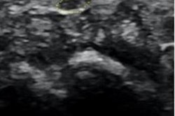
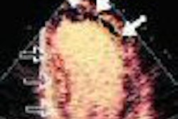
One radiologist imaged 206 wrists in the patients using a 7- to 15-MHz linear-array transducer on an HDI 5000 scanner (Philips Medical Systems, Andover, MA). Grayscale sonography was used to evaluate the presence of median nerve swelling, edema, and flattening and increased bowing of the flexor retinaculum, while color Doppler sonography was employed to evaluate the presence of nerve hypervascularization.
The researchers compared the sensitivity and specificity of each sonographic feature with nerve conduction studies. Carpal tunnel syndrome was confirmed by nerve conduction studies in 172 wrists.
Grayscale sonography revealed at least one abnormal finding in 181 wrists, while color Doppler sonography depicted intraneural blood vessels in 174 wrists and correctly identified carpal tunnel syndrome in 164, according to the researchers. All five sonography criteria were present in 65 wrists (32%).
"Of 172 wrists with carpal tunnel syndrome, 11 met only one sonography criterion for carpal tunnel syndrome -- that is, median nerve hypervascularization in seven wrists, nerve swelling in three, and nerve flattening in one," the authors wrote.
In 153 wrists (89%) with carpal tunnel syndrome, both nerve swelling and nerve hypervascularization were seen, while 15 wrists (9%) displayed either nerve swelling or nerve hypervascularization. In cases of disagreement between nerve swelling and nerve hypervascularization, nerve hypervascularization correctly correlated with nerve conduction studies in 11 wrists, and nerve swelling correlated correctly in four cases, according to the study team.
Nerve swelling turned in the highest accuracy (91%) among the grayscale sonography criteria, while the presence of intraneural hypervascularization showed the highest accuracy (95%) among all sonography criteria, according to the researchers. From logistic regression analysis, the researchers found that nerve hypervascularization was the only variable that independently predicted median nerve entrapment (odds ratio 16.4; 95% confidence interval).
"Color Doppler sonography contributed more than grayscale sonography to the perception of the circulatory status of the median nerve and consequently to the advantageous characterization of nerve involvement in patients with clinically suspected carpal tunnel syndrome," the authors concluded. "This could have a considerable impact on the early diagnosis of median nerve compression and on patient selection for carpal tunnel decompression."
By Erik L. Ridley
AuntMinnie.com staff writer
May 18, 2006
Related Reading
Ergonomics -- It's all about you, November 23, 2005
OMT for carpal tunnel no better than placebo ultrasound, November 9, 2004
Tackling ergonomic issues in sonography, September 13, 2004
Sonography useful in diagnosing carpal tunnel syndrome, July 5, 2004
Work-related musculoskeletal injuries continue to plague rads, August 20, 2003
Copyright © 2006 AuntMinnie.com