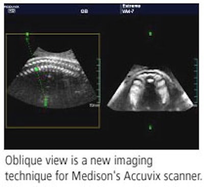
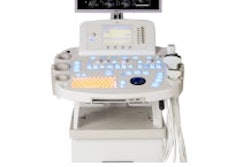
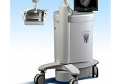
Medison of Cypress, CA, will present new features to Accuvix, the company's flagship ultrasound system that supports 2D, 3D, and 4D scanning.

New to Accuvix is a 3D toolset called 3D XI, which includes techniques such as 3D multislice imaging, oblique view, cross volume, cube volume, and dynamic MR.
The 3D multislice imaging technique displays the acquired 3D anatomy as sequential images, similar to the display of CT and MR scans. It also provides documentation of anatomy and pathology through the sequential display format. The user is able to select the slice thickness, from 0.5 mm to 5 mm. The CT/ MR-like display format is designed for increased comprehension of the acquired anatomy within the 3D dataset.
The 3D multislice technology provides more efficient scan time when performing ultrasound examinations, according to Medison. The ability to acquire the anatomy of interest in as fast as two seconds using the automatic acquisition 3D transducer, in combination with the instantaneous display of multislice imaging, is intended to decrease sonographer injury often associated with the repetitive motion of conventional 2D scanning.
The oblique view technique provides the exact corresponding cross-sectional view with the turn of a dial, while cross volume displays the 3D multiplanar anatomy as three intersecting scan planes, which can be rotated, providing improved comprehension of the spatial relationships of anatomical planar images.
With cube volume imaging, 3D planar images are presented in a cube format, providing an understanding of the spatial relationships between the 3D planar images, according to the company. Finally, dynamic MR is designed to remove the traditional speckle pattern from the ultrasound image, resulting in superior contrast resolution and increased visualization of anatomical borders.
By Wayne Forrest
AuntMinnie.com contributing writer
November 9, 2005
Copyright © 2005 AuntMinnie.com







![Examples of ultrasound findings and techniques. (A) Images in a 39-year-old male patient with a mass in the left thigh. The mass is heterogeneous on the B-mode US image (compared with the patient in D) and showed increased microvascularity (superb microvascular imaging [SMI]) and shear-wave elastography (SWE) values. Undifferentiated pleomorphic sarcoma was diagnosed at biopsy (with pleomorphic rhabdomyosarcoma in surgical specimen). (B) Images in an 18-year-old male patient with a mass in the left leg. The mass is hypoechoic on the B-mode image, with no other findings suggestive of malignancy. The lesion is in contact with the cortex of the tibia, which is slightly irregular. CT revealed a doubtful anteromedial tibial erosion. The microvascular study demonstrated high vascularization, suggestive of malignancy. Periosteal Ewing sarcoma was diagnosed with both histologic and immunohistochemical confirmation. (C) Images in a 69-year-old female patient with a lump growing on the outside of the left leg. Multiple SWE examinations were performed (please note the high values obtained in the measurements, whereas the color map highlights the stiffness relative to adjacent tissues). SMI showed areas of increased vascularization to target for sampling. Undifferentiated spindle cell sarcoma was diagnosed at biopsy, with residual leiomyosarcoma in the surgical specimen after neoadjuvant therapy. (D) Images in a 56-year-old female patient with a mass in the right thigh. The mass is heterogeneous at both B-mode ultrasound (similar to patient A) and MRI (coronal T2-weighted spectral attenuated inversion recovery [SPAIR]; T1-weighted pre-contrast and postcontrast imaging), which even shows uptake after the administration of paramagnetic contrast material, which is traditionally suggestive of malignancy. Low values at SMI and elastography are suggestive of benignity. Spindle cell lipoma was diagnosed at biopsy, with atypical spindle cell lipomatous tumor in the surgical specimen.](https://img.auntminnie.com/mindful/smg/workspaces/default/uploads/2026/08/images-radiol250278fig2.APCFLSvX6p.jpg?auto=format%2Ccompress&fit=crop&h=112&q=70&w=112)