AUSTIN, TX -- Microflow imaging performs just as well as contrast-enhanced ultrasound (CEUS) for guiding tumor ablation and may be an economical alternative, according to research presented April 8 at UltraCon.
In her talk, Ziyue Hu, MD, from Sichuan Cancer Hospital in China highlighted findings showing that along with yielding comparable performance in treating thyroid nodules, microflow imaging in this area is less costly and invasive than CEUS.
“We found that in the microwave ablation procedure for thyroid nodules that … microflow imaging offers the advantages of reducing surgical expenses, which lessens the economic burden for patients,” Hu said.
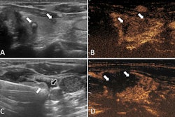
Ultrasound-guided microwave ablation determines complete necrosis of targeted tumor areas, with CEUS being able to observe intra-tissue blood flow perfusion. This, in turn, allows for sensitive monitoring of residual lesions.
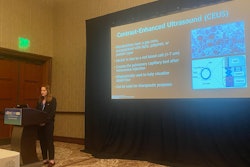
 Ziyue Hu, MD, from Sichuan Cancer Hospital in China highlighted findings at UltraCon, showing that along with comparable performance in treating thyroid nodules, microflow imaging is a less costly and invasive approach to tumor ablation than CEUS.Amerigo Allegretto
However, Hu said that CEUS has its share of limitations, including being an invasive and costly tool and being inaccessible for patients with contrast allergies or cardiopulmonary dysfunction.
Microflow imaging meanwhile is an ultrasound method that visualizes small vessels and slow blood flow signals without using contrast agents. This method is noninvasive and enables real-time, sensitive evaluation of target areas, according to Hu. She and colleagues compared the effectiveness of microflow imaging and CEUS during microwave ablation of thyroid nodules.
The study included 88 thyroid nodules in 73 patients. Of the total, 38 patients with 43 thyroid nodules underwent microflow imaging guidance while 35 patients with 45 nodules underwent CEUS guidance. The study also evaluated patients at several follow-up points within an 18-month period.
The researchers found that after more than one year of follow-up, both patient groups experienced complete thyroid nodule treatment. Additionally, there were no significant differences in volume and volume reduction rate between both groups.
| Comparison in volume, volume reduction rate between microflow imaging, CEUS | |||||||
|---|---|---|---|---|---|---|---|
| Volume (ml) | Volume reduction rate | ||||||
| Follow-up time | CEUS | Microflow imaging | p-value | CEUS | Microflow imaging | p-value | |
| 1 month | 1.97 | 2.12 | 0.318 | 0.52 | 0.52 | 0.208 | |
| 3 months | 1.26 | 0.89 | 0.986 | 0.72 | 0.71 | 0.269 | |
| 6 months | 0.58 | 0.44 | 0.414 | 0.84 | 0.85 | 0.892 | |
| 12 months | 0.53 | 0.25 | 0.216 | 0.91 | 0.93 | 0.255 | |
| 18 months | 0.27 | 0.11 | 0.285 | 0.92 | 0.96 | 0.231 | |
While both groups experienced similar supplementary ablation times, procedure times, and complications from pain and edema, Hu highlighted that microflow imaging had significantly less financial cost than CEUS. This included an average of 11,334.36 yuan ($1,567.49 U.S.) versus 12,948.17 yuan ($1,790.67, p = 0.041).
"The difference in cost ... is mainly due to the varying amounts of contrast agent used," Hu said. "When a patient has multiple thyroid nodules or needs immediate supplemental ablation during microwave ablation, multiple CEUS examinations will be conducted. This leads to a larger amount of contrast agent being used than usual, which results in higher costs."
She added that during a retrospective examination of the results, the team observed pronounced gas echo generated immediately after ablation can impede microflow imaging. She said with this in mind, it is important for treatment teams to allow time for gas to disperse before undertaking evaluations.