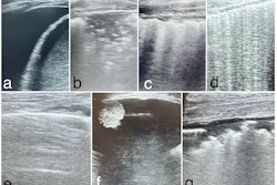
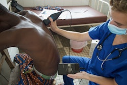
Some of the top "domains" of point-of-care ultrasound (POCUS) incorporate the technology into lung ultrasound applications, according to research published in the Journal of Radiology Nursing.
A team led by the POCUS Certification Academy, part of Inteleos in Rockville, MD, published a list of the top 25 POCUS community-defined practice domains, dubbed “POCUS25.” They found via survey data that the POCUS community believes the most important domains are tied to imaging of the pneumothorax, pericardial effusion and cardiac tamponade detection, normal lung ultrasound, basic practical physics, and extended focused assessment with sonography in trauma (eFAST).
“It’s exciting to see the POCUS community help put this list together,” said corresponding author Victor Rao, MBBS, from the academy to AuntMinnie.com.
As POCUS continues to grow in popularity inside the clinic, there is a need for standardized training and best practices. This includes establishing definitions for core competencies that span multiple specialties.
The academy researchers addressed this in the POCUS25 study, a global end-user study that aimed to identify the top 25 practice domains. They reasoned that with POCUS being a “global grassroots movement” led by end-users, an expert panel would not be likely to represent global trends and practices.
Academy director Jasmine Rockett said the study stemmed from POCUS users seeking guidance and understanding on the proficient use of POCUS. Study co-author R. Eugene Zierler, MD, added that understanding how and where POCUS is primarily used helps better inform POCUS education and certification structures.
The researchers included data from 360 participants. Of the participants who filled out survey data regarding gender and age, 58% were male and 46% were over 45 years old. And while most participants were based in the U.S. (60%), other countries represented in the survey included Canada, Australia, Mexico, India, Brazil, Pakistan, the U.K., and others.
Additionally, 50% of respondents were physicians while 23% were sonographers. Specialties represented included emergency medicine (19%), family medicine (12%), radiology (10%), and ob/gyn (9%). Other specialties included cardiology, internal medicine, and anesthesiology, among several others.
Furthermore, 38% of respondents reported having an advanced level of POCUS use while 35% reported intermediate skill levels.
To gather their ranked list, the researchers asked the participants how important it is for an entry-level practitioner to know the POCUS practice domains. They applied a weighted average to these “importance” responses.
The following are the top 10 practice domains as listed in the POCUS25 list:
- Pneumothorax – B-mode and M-mode
- Pericardial effusion and cardiac tamponade detection and differentiation from pleural effusion; assess inferior vena cava to support cardiac tamponade findings
- Normal lung ultrasound
- Basic practical ultrasound physics and knobology
- Protocol – eFAST
- Focused cardiac ultrasound (FoCUS)
- Gross estimation of left and right ventricular systolic function
- Extremes of volume status
- Abdominal aortic aneurysm (AAA) screening
- Deep vein thrombosis
Rao said he was most surprised by lung ultrasound being among the top practice domains given its recent acceptance into the ultrasound field.
The results indicate that while the POCUS25 practice domains are relevant to almost all medical specialties, the top domains are in acute care settings such as emergency medicine, trauma, and critical care, among others. This reflects the 19% of respondents being emergency medicine physicians. The lower half of the list includes the ob/gyn practice domains and domains for general abdominal, soft tissue, and musculoskeletal ultrasound.
The researchers highlighted that such findings could encourage an increased use of ultrasound to improve and accelerate patient care in the future. They added that ongoing research is needed to identify new emerging trends and maintain a globally relevant POCUS core competency list. One future consideration they noted was the role and use of AI in POCUS.
“We are collecting data even beyond this point. POCUS is evolving, and we don’t want it to become redundant and out of context a few years down the road,” told AuntMinnie.com. “It’s designed to run forever, actually.”
Rockett echoed that this study is a starting point, adding that it will be important to continue collaborating with the POCUS community to continue establishing resources and education protocols.
The full list can be found here.