CHICAGO - Ultrasound-guided hydrodissection is an effective treatment for carpal tunnel syndrome, according to research results presented November 30 at RSNA meeting.
A team led by Dr. Anupama Tandon of the University College of Medical Sciences in Delhi, India, and colleagues found that at six-month follow-up, hydrodissection delivered complete and long-term relief to patients.
"It came as a pleasant surprise when this simple procedure of ultrasound-guided hydrodissection provided patients with long-term relief," Tandon said in an RSNA statement.
Carpal tunnel syndrome is the most common and widely known form of entrapment neuropathy, affecting about 3% of the U.S. population. Steroid injections and surgery are often required to treat the condition, with most common surgical method involving cutting the carpal ligament to reduce pressure on the median nerve.
Hydrodissection is a less invasive alternative, involving the injection of a liquid -- usually saline -- into the nerve to separate it from the surrounding tissue. Ultrasound is used during the procedure to accurately identify nerves.
Tandon and colleagues sought to assess the efficacy of hydrodissection for carpal tunnel syndrome treatment via a study that included 63 patients. Participants were divided into three groups: One which received ultrasound-guided hydrodissection with just a saline injection; a second which received ultrasound-guided hydrodissection with an injection mixture of saline and corticosteroid; and a third which received just an ultrasound-guided corticosteroid injection with no hydrodissection. The team followed up with patients at four weeks, 12 weeks, and six months post-procedure.
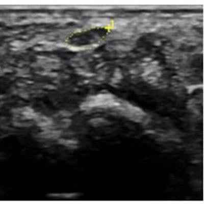
Ultrasound image of median nerve before and after hydrodissection procedure in a patient with carpal tunnel syndrome. This case had cross-sectional area of 12 mm2 pre procedure that reduced to 7 mm2 post procedure (dashed yellow lines depicting the median nerve). Image courtesy of RSNA and Dr. Anupama Tandon.All three groups of patients showed a reduction in pain at the four-week mark. By the 12-week and six-month mark, both groups that received ultrasound-guided hydrodissection showed further improvement, while the group that received just a corticosteroid injection reported a recurrence of symptoms. In addition, follow-up ultrasounds showed a significant reduction of median nerve cross-sectional area in both hydrodissection groups, with group one demonstrating a reduction of 43% and group two of 46%. Group three showed only an 11% reduction.
"The procedure is short, requiring only 10 to 15 minutes, and is cost-effective, since it doesn't require any high-end equipment," Tandon concluded.


![Examples of ultrasound findings and techniques. (A) Images in a 39-year-old male patient with a mass in the left thigh. The mass is heterogeneous on the B-mode US image (compared with the patient in D) and showed increased microvascularity (superb microvascular imaging [SMI]) and shear-wave elastography (SWE) values. Undifferentiated pleomorphic sarcoma was diagnosed at biopsy (with pleomorphic rhabdomyosarcoma in surgical specimen). (B) Images in an 18-year-old male patient with a mass in the left leg. The mass is hypoechoic on the B-mode image, with no other findings suggestive of malignancy. The lesion is in contact with the cortex of the tibia, which is slightly irregular. CT revealed a doubtful anteromedial tibial erosion. The microvascular study demonstrated high vascularization, suggestive of malignancy. Periosteal Ewing sarcoma was diagnosed with both histologic and immunohistochemical confirmation. (C) Images in a 69-year-old female patient with a lump growing on the outside of the left leg. Multiple SWE examinations were performed (please note the high values obtained in the measurements, whereas the color map highlights the stiffness relative to adjacent tissues). SMI showed areas of increased vascularization to target for sampling. Undifferentiated spindle cell sarcoma was diagnosed at biopsy, with residual leiomyosarcoma in the surgical specimen after neoadjuvant therapy. (D) Images in a 56-year-old female patient with a mass in the right thigh. The mass is heterogeneous at both B-mode ultrasound (similar to patient A) and MRI (coronal T2-weighted spectral attenuated inversion recovery [SPAIR]; T1-weighted pre-contrast and postcontrast imaging), which even shows uptake after the administration of paramagnetic contrast material, which is traditionally suggestive of malignancy. Low values at SMI and elastography are suggestive of benignity. Spindle cell lipoma was diagnosed at biopsy, with atypical spindle cell lipomatous tumor in the surgical specimen.](https://img.auntminnie.com/mindful/smg/workspaces/default/uploads/2026/08/images-radiol250278fig2.APCFLSvX6p.jpg?auto=format%2Ccompress&fit=crop&h=100&q=70&w=100)


![Examples of ultrasound findings and techniques. (A) Images in a 39-year-old male patient with a mass in the left thigh. The mass is heterogeneous on the B-mode US image (compared with the patient in D) and showed increased microvascularity (superb microvascular imaging [SMI]) and shear-wave elastography (SWE) values. Undifferentiated pleomorphic sarcoma was diagnosed at biopsy (with pleomorphic rhabdomyosarcoma in surgical specimen). (B) Images in an 18-year-old male patient with a mass in the left leg. The mass is hypoechoic on the B-mode image, with no other findings suggestive of malignancy. The lesion is in contact with the cortex of the tibia, which is slightly irregular. CT revealed a doubtful anteromedial tibial erosion. The microvascular study demonstrated high vascularization, suggestive of malignancy. Periosteal Ewing sarcoma was diagnosed with both histologic and immunohistochemical confirmation. (C) Images in a 69-year-old female patient with a lump growing on the outside of the left leg. Multiple SWE examinations were performed (please note the high values obtained in the measurements, whereas the color map highlights the stiffness relative to adjacent tissues). SMI showed areas of increased vascularization to target for sampling. Undifferentiated spindle cell sarcoma was diagnosed at biopsy, with residual leiomyosarcoma in the surgical specimen after neoadjuvant therapy. (D) Images in a 56-year-old female patient with a mass in the right thigh. The mass is heterogeneous at both B-mode ultrasound (similar to patient A) and MRI (coronal T2-weighted spectral attenuated inversion recovery [SPAIR]; T1-weighted pre-contrast and postcontrast imaging), which even shows uptake after the administration of paramagnetic contrast material, which is traditionally suggestive of malignancy. Low values at SMI and elastography are suggestive of benignity. Spindle cell lipoma was diagnosed at biopsy, with atypical spindle cell lipomatous tumor in the surgical specimen.](https://img.auntminnie.com/mindful/smg/workspaces/default/uploads/2026/08/images-radiol250278fig2.APCFLSvX6p.jpg?auto=format%2Ccompress&fit=crop&h=112&q=70&w=112)