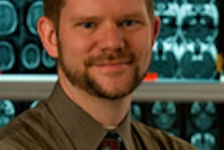
Like many other imaging modalities, ultrasound has experienced significant technology changes -- and workflow modifications. But adoption of digital image management technology can improve productivity, aiding users in handling the new workflow requirements, according to Dr. Steven Horii of the University of Pennsylvania Medical Center (UPMC) in Philadelphia.
PACS and digital healthcare and information management systems (DHIMS) offer "a distinct advantage for productivity gains and may also help with detractors from productivity, such as intraoperative ultrasound," Horii said.
He spoke at the PACS 2007 conference in San Antonio, sponsored by the University of Rochester School of Medicine & Dentistry in Rochester, NY.
Ultrasound practices are in the midst of change. Many ultrasound practices are now filmless. And color flow Doppler is used in many examinations, which means that the images include color information. Volumetric imaging is also on the rise, and instruments and transducers are increasing in variety and capability, Horii said.
Ultrasound studies are also bigger than they used to be -- color images feature 24-bit pixels, and image matrices are larger (1024 x 768 instead of 640 x 480) as well, he said.
In addition, the number of images is growing. At UPMC, the number of ultrasound images per exam increased from 30 to 46.5 over an 18-month period between 2004 and 2006, Horii said. That data didn't include cine loops, which were counted as one image.
The use of cine loops is increasing due to volumetric scanning for 3D and 4D imaging. And ultrasound is also finding increased utilization for guiding biopsies, in interventional radiology for procedure guidance, and in the OR for surgical guidance, Horii said.
Workflow changes
Changes in ultrasound have also resulted in new workflow. Shifting from film cassettes to centralized printing saved three to five minutes per patient per sonographer at UPMC, while a change to PACS yielded slight time savings over centralized printing, Horii said. Adopting PACS along with DICOM modality worklist saved another one to two minutes per patient per sonographer and reduced data entry errors.
At UPMC, the ultrasound section examination volume has been growing at approximately 10% per year. During that time, the institution has increased its number of sonographers by only one full-time employee (a 10% increase), while increasing its number of examination rooms from eight to 10.
"We are being more productive," Horii said.
Information systems have been key in improving productivity, according to Horii. Prior examinations are readily available from PACS, and reports are available from the RIS, allowing sonographers to focus their examinations.
"All of our sonographers know that if the patients have had a prior study, they should look at those reports," he said. "They're very good. They also look at the CT reports; they look at the MR reports to see what was found and what it is that they can direct their exam to find out."
DICOM modality worklist obviates data entry, and the RIS supports some automation of managing patient transport, he said.
A general PACS allows sonographers and radiologists to perform examination correlation, while a Web-based physician portal enables retrieval of laboratory, pathology, and consult results, Horii said. Library and search engines such as Yottalook (iVirtuoso, Baltimore) provide for just-in-time diagnostic support.
In changes to reporting, Horii has started inserting Web and other references in reports. At present, however, the RIS does not support maintaining the Web links if the report is viewed online, he said.
Separate ultrasound PACS
Many sites have used a separate ultrasound PACS, as only relatively recently have PACS vendors started supporting the tools needed for viewing and interpreting ultrasound examinations, Horii said.
To integrate ultrasound into a DHIMS, institutions can choose from moving to a single PACS or keeping a separate PACS but combining storage, he said. UPMC is using the latter approach.
"All of our image acquisition is done into our ultrasound miniPACS and then it gets shipped off into our main hospital PACS," Horii said.
With the separate PACS method, users will have to select either a push model (where the ultrasound PACS initiates a DICOM send action when the exam is complete) or a pull model (where the general PACS performs a DICOM query/retrieve action when it's informed of the study completion by the RIS).
The common storage approach also requires that patient demographic and exam information match in both the ultrasound PACS and general PACS databases, Horii said.
"The use of DICOM modality worklist helps a lot because it really ensures that happens," he said.
This model also serves as a backup for the long-term storage, Horii said. Studies are stored on both the general PACS storage system and a separate system used by the ultrasound PACS.
Intraoperative ultrasound
Intraoperative ultrasound can also offer a workflow challenge. Nonscheduled studies can be a complex process to handle, involving steps carried out by several people, Horii said. Ultrasound schedules have to be adjusted, and surgeons and patients are also impacted.
For ultrasound, the problem is the break in workflow. Sonographers have to rearrange their workflow, and the radiologist has to stop reading cases or teaching activities to handle the unscheduled exams, Horii said.
Information systems and technology can help with scheduling, as well as seeing and moving images, he said.
Predictive scheduling can assist, as most intraoperative ultrasound tends to be for specific types of cases. Scheduling systems can be aware of OR schedules days, if not weeks, in advance and can flag possible intraoperative ultrasound cases and alert radiology, Horii said. They also make it easy for the surgeon to schedule ultrasound ahead of time.
An alert system in the surgical workflow manager can also help surgeons notify radiology staff closer to the time when they're needed, Horii said.
As for viewing of images, a display system with a flat-panel display on a boom could help, he said. Most ultrasound machines have a video output connector, while newer ones have a video graphics array (VGA) or digital visual interface (DVI) connector. "These could be used to drive a multipurpose display in the OR," Horii said.
A network connection in the OR that can be used by the ultrasound machines could also be useful.
"It may be a challenge to get to the ultrasound PACS from the OR network, but this is where IT infrastructure design can help," Horii said.
By Erik L. Ridley
AuntMinnie.com staff writer
April 23, 2007
Related Reading
RIS-SR-PACS combo speeds radiology reporting, April 17, 2007
Norwegian teleradiology project overcomes technical, political hurdles, April 11, 2007
3D ultrasound offers efficiency improvements, March 29, 2007
Planning for PACS downtime eases impact on operations, March 22, 2007
RIS/PACS provides potential for performance, safety improvements, March 21, 2007
Can ultrasound benefit from the rest of radiology?, March 8, 2007
Copyright © 2007 AuntMinnie.com