BERLIN - One-third of cardiac resynchronization therapy (CRT) procedures in patients with congestive heart failure (CHF) experience hemodynamic failure, while another 12% are marred by implantation failure. MR coronary vein imaging holds promise for preprocedural assessment of the veins in CHF patients, according to a presentation Monday at the International Society for Magnetic Resonance in Medicine (ISMRM) meeting.
"CRT is achieved by simultaneously pacing the right ventricle and the left ventricle to relieve CHF symptoms. Transvenous implantation is done through the coronary sinus using an available lateral vein branch," explained Dr. Reza Nezafat from Beth Israel Deaconess Medical Center in Boston. Nezafat's colleagues are from Boston-based Harvard Medical School and Philips Research North America in New York City.
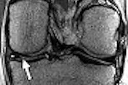
Currently, vein assessment is done either with invasive x-ray fluoroscopy or iodinated contrast-enhanced CT venography, Nezafat said. "The problem with (CT venography) is that it requires a high radiation dose and it requires iodinated contrast. In this patient population, iodinated contrast may be problematic because these patients also have renal insufficiency," he explained.
Nezafat outlined the MR coronary vein imaging technique that his group developed as an alternative imaging method. First, for acceptable tissue contrast, they determined that magnetization transfer preparation offered several advantages over no preparation or T2 magnetization preparation. Magnetization transfer preparation created contrast between blood and myocardium by suppressing the myocardium but not the veins, he stated. Adding a Gaussian-sinc pulse shape also resulted in the lowest specific absorption rate and less artifact, he added.
In terms of the imaging sequence, the authors ruled out steady-state free precession (SSFP). "SSFP is not suited for (coronary vein imaging) because we're imaging through the transit of SSFP," Nezafat said. "If we are imaging during that time, the SSFP signal is proportional to T2 if we are using the high flip angle. But we're interested in imaging the veins, and those have lower T2s so the high flip angle would not be an option."
Instead, they found a gradient-recalled echo (GRE) sequence, along with the magnetization transfer preparation, produced less contrast-to-noise and signal-to-noise ratios between the veins and myocardium.
Finally, with regard to trigger delay, they preferred imaging during the systolic time versus diastolic time. "The coronary sinus and vein size change through the cardiac cycle, mainly due to elasticity," he said. "We see a larger coronary sinus cross-section systolically. The end systolic time overlaps with the maximum size of the vein, which is when imaging should be done."
The pre-CRT protocol they have devised starts with MR coronary vein imaging, followed by cardiac function testing, dysynchrony with tagging, and a viability scan, Nezafat said. Using this method, they can evaluate variations in vein anatomy, as well as the size of the branches and angles for the CRT leads. More important, MR coronary vein imaging can pick out the "unfavorable veins," he said. These patients should then go directly to epicardial surgical implantation.
By Shalmali Pal
AuntMinnie.com staff writer
May 22, 2007
Related Reading
Benefit of cardiac resynchronization therapy predicted by preimplant BNP, February 2, 2007
Cardiac resynchronization attenuates mitral regurgitation, August 7, 2006
QRS duration tied to cardiac resynchronization outcome, June 28, 2005
Cardiac resynchronization therapy sharply improves survival in heart failure, March 8, 2005
Tissue synchronization imaging predicts outcome of cardiac resynchronization, March 7, 2005
Copyright © 2007 AuntMinnie.com