Structural MRI measures can predict fatigue severity in individuals with multiple sclerosis, researchers have found.
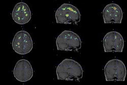
MR imaging of more than 4,000 patients showed that baseline lesion burden and lower whole-brain volumes were linked to multiple sclerosis fatigue, wrote a team led by Alexandra Simpson, MD, of Johns Hopkins University in Baltimore, MD. The study results were published March 25 in Multiple Sclerosis.
"Higher baseline brain parenchymal and lower T2 lesion volume at baseline [imaging] were associated with lower odds of subsequent periods of elevated fatigue," the group reported.
Findings from previous research on radiological markers of multiple sclerosis fatigue have been conflicting, Simpson and colleagues explained. To clarify the issue, they conducted a study that explored any associations of lesion and brain compartment volumes with fatigue severity and persistence in people with the condition.
The study included 4,012 participants with multiple sclerosis who had varying levels of fatigue: 2,058 had no fatigue, 629 had mild fatigue, and 1,325 had moderate-to-severe fatigue. The researchers used data from the Multiple Sclerosis Partners Advancing Technology and Health Solutions (MS PATHS) network, and they tracked MRI multiple sclerosis fatigue predictors such as baseline brain parenchymal and gray matter fractions and T2 lesion volume. They analyzed these predictors using the Quality of Life in Neurological Disorders (Neuro-QOL) fatigue subscore, with elevated fatigue symptoms defined as T-score > mean + 0.5 standard deviation over follow-up.
The group found the following:
- One standard deviation greater baseline brain parenchymal and gray matter fractions were associated with 0.83 (p < 0.001) and 0.38 (p = 0.02) lower values in the baseline Neuro-QOL fatigue T-score.
- A one standard deviation lower measure of total T2 lesion volume was associated with a 0.49 (p < 0.001) lower baseline fatigue T-score.
- Higher brain parenchymal and lower T2 lesion volume at baseline were associated with lower odds of subsequent periods of elevated fatigue.
"Baseline lesion burden and lower generalized whole-brain volumes were associated with MS fatigue in cross-sectional and longitudinal analyses in a large, real-world cohort of people with multiple sclerosis," Simpson and colleagues concluded.
The complete study can be found here.


![Overview of the study design. (A) The fully automated deep learning framework was developed to estimate body composition (BC) (defined as subcutaneous adipose tissue [SAT] in liters; visceral adipose tissue [VAT] in liters; skeletal muscle [SM] in liters; SM fat fraction [SMFF] as a percentage; and intramuscular adipose tissue [IMAT] in deciliters) from MRI. The fully automated framework comprised one model (model 1) to quantify different BC measures (SAT, VAT, SM, SMFF, and IMAT) as three-dimensional (3D) measures from whole-body MRI scans. The second model (model 2) was trained to identify standardized anatomic landmarks along the craniocaudal body axis (z coordinate field), which allowed for subdividing the whole-body measures into different subregions typically examined on clinical routine MRI scans (chest, abdomen, and pelvis). (B) BC was quantified from whole-body MRI in over 66,000 individuals from two large population-based cohort studies, the UK Biobank (UKB) (36,317 individuals) and the German National Cohort (NAKO) (30,291 individuals). Bar graphs show age distribution by sex and cohort. BMI = body mass index. (C) After the performance assessment of the fully automated framework, the change in BC measures, distributions, and profiles across age decades were investigated. Age-, sex-, and height-adjusted body composition reference curves were calculated and made publicly available in a web-based z-score calculator (https://circ-ml.github.io).](https://img.auntminnie.com/mindful/smg/workspaces/default/uploads/2026/05/body-comp.XgAjTfPj1W.jpg?auto=format%2Ccompress&fit=crop&h=100&q=70&w=100)


![Overview of the study design. (A) The fully automated deep learning framework was developed to estimate body composition (BC) (defined as subcutaneous adipose tissue [SAT] in liters; visceral adipose tissue [VAT] in liters; skeletal muscle [SM] in liters; SM fat fraction [SMFF] as a percentage; and intramuscular adipose tissue [IMAT] in deciliters) from MRI. The fully automated framework comprised one model (model 1) to quantify different BC measures (SAT, VAT, SM, SMFF, and IMAT) as three-dimensional (3D) measures from whole-body MRI scans. The second model (model 2) was trained to identify standardized anatomic landmarks along the craniocaudal body axis (z coordinate field), which allowed for subdividing the whole-body measures into different subregions typically examined on clinical routine MRI scans (chest, abdomen, and pelvis). (B) BC was quantified from whole-body MRI in over 66,000 individuals from two large population-based cohort studies, the UK Biobank (UKB) (36,317 individuals) and the German National Cohort (NAKO) (30,291 individuals). Bar graphs show age distribution by sex and cohort. BMI = body mass index. (C) After the performance assessment of the fully automated framework, the change in BC measures, distributions, and profiles across age decades were investigated. Age-, sex-, and height-adjusted body composition reference curves were calculated and made publicly available in a web-based z-score calculator (https://circ-ml.github.io).](https://img.auntminnie.com/mindful/smg/workspaces/default/uploads/2026/05/body-comp.XgAjTfPj1W.jpg?auto=format%2Ccompress&fit=crop&h=112&q=70&w=112)


