Imagine a radiology department outfitted with the latest innovations in workstations, PACS, RIS, computer-aided detection (CAD), and advanced image visualization tools, all adhering to Integrating the Healthcare Enterprise (IHE) profiles. For its meeting in Vienna this week, the European Congress of Radiology (ECR) has assembled such a collection of products for its Imagine project, which promises a preview of the radiology department of the near future.
The Imagine project is not a virtual hospital, but rather an intelligent department, according to Bart ter Haar Romeny, Ph.D., one of the project organizers and a professor of biomedical image analysis at the Eindhoven University of Technology in the Netherlands.
"We have not connected all exhibits, but we have integrated a few of them to six IHE locations," ter Haar Romeny said. "And at each location, a modality reading room is simulated."
Featured radiological applications in the Imagine project include CAD, image analysis, interactive and virtual-reality 3D displays, database retrieval, PACS, and image-guided surgery planning, he said.
"Imagine shows the intelligent department we foresee in the near future. The real intelligence goes beyond merely connecting things; it is also in the intelligence of the applications," ter Haar Romeny said.
The Imagine project will mimic a real imaging department, but conventional reading rooms will be replaced by rooms for specific functions such as CAD, image processing, and RIS-PACS integration. Imagine will have eight rooms situated in the ECR entrance matrix, where experts can meet with interested visitors for small group discussions.
Visitors will be met at an admission desk, and will receive a report of their visit when they leave. Guided tours will be offered three times each day, according to project organizers.
A collection of research institutes, universities, equipment, and software vendors will showcase their latest offerings at the conference.
AngioVis
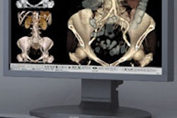
This exhibit will illustrate visualization tools for CT angiography (CTA) developed by the Angiographic Visualization Project at the Vienna University of Technology in Austria. The group is working on a diagnosis tool for detecting and classifying arterial disease in routine clinical use. Several interconnected topics, ranging from clinical background and image acquisition techniques to final image evaluation and interpretation, will be part of this interdisciplinary exhibit.
Austrian Medical Imaging Processing Group of Vienna, Graz, and Innsbruck, Austria
This collaborative team will be spotlighting a clutch of new technologies as its part of the Imagine project:
B-ROB II is a robotic system for percutaneous interventions to assist ultrasound- and CT-guided percutaneous interventions, such as biopsies, and to provide the physician with a needle-guidance tool that enables access to difficult-to-reach lesions.
Unified patient is a Web-based software and medical media database supporting surgical planning and interdisciplinary meetings, including multimedia storage for medical imaging data and patient records.
CAPPA IRAD is an immobilization and registration device for image-guided percutaneous interventions. In addition, registration algorithms for accurate fusion of various preoperative imaging modalities such as CT, MR, SPECT, PET, and ultrasound will be demonstrated.
A virtual liver planning system, developed using cross-sectional imaging from CT and MRI, will utilize a head-mounted display to provide visualization of anatomy and pathology.
A patient-specific computational analysis of transvenous defibrillation will assess the predictive capacity of computer models by comparing the results of patient-specific simulations to clinical defibrillation thresholds.
A CAD rheumatoid arthritis application will offer an interactive automated quantification of bone lesions, and an automated estimation of the bone contour from serial hand radiographs in patients with rheumatoid arthritis.
A fused deposition model (FDM) project for operation planning will be demonstrated. The project matches functional and anatomical data to create a plastic model that can be used by surgeons to plan access and operation techniques.
Barco (Voxar)
Edinburgh, U.K.-based Barco will present its latest multiplanar reformatting (MPR), 3D toolset, and specialized clinical application modules designed for a PACS environment. The company said it will also demonstrate performance advances made in software- and hardware-based 3D rendering using high-performance graphics accelerators.
Biomedical Imaging Lab, Agency for Science, Technology and Research, Singapore
This group will be showcasing its atlas-assisted analysis of brain scans. The team believes that model-enhanced radiology may speed up interpretation of brain scans while flattening the learning curve for radiologists. The exhibit contains three computer demos: rapid and automatic atlas-based interpretation of MRI normal brain scans, assistance in interpreting stroke images by means of anatomical and blood supply territories atlases, and atlas-assisted interpretation of PET/SPECT brain images.
A Chinese-language version of the Cerefy Atlas of Brain Anatomy work-in-progress will also be exhibited, according to the group.
Charité - Unversitätsmedizin, Campus Virchow-Klinikum, Berlin
The university-based researchers will be demonstrating Marvin, a modular image archive, and a remote data entry (RDE) system, Asena, as part of the ECR Imagine project.
The Marvin application offers an Everything Online (EOL) DICOM image archive with an integrated Web interface for image retrieval. The Asena tool allows users to set up customized Web-based applications for RDE. The combination of applications enables digital image archiving and remote data collection, including secure exchange of DICOM images, according to the researchers.
Computer Graphics Lab, ETH, Co-Me, Zurich, Switzerland
The research group will be spotlighting its Towards Hysteroscopy Simulation: Interactive Simulation Components for Deformable Tetrahedral Meshes. Their work focuses on developing interactive methods and algorithms for surgery simulation and training, investigating approaches to tetrahedral mesh generation, deformable modeling, deformable collision handling, fluid simulation, and the simulation of surgical instruments.
The Cairo Montenotte, Italy-based developer will be highlighting interoperability for integrated imaging and information systems, and demonstrating these capabilities in its LifeWeb suite.
The vendor will show how a large enterprise PACS, made by multiple, heterogeneous data repositories, can be arranged as a single virtual collaborative system. The firm will also exhibit the implementation of data coding that allows cross-enterprise data portability, including images as well as report and diagnostic documentation.
In addition, the company will demonstrate the use of secure Web technology to distribute and visualize images in a heterogeneous network environment comprised of a local intranet, the Internet, and wireless network.
Interdisciplinary Cardiac Imaging Center, Department of Radiology, Graz, Austria
The research group will demonstrate the results of a multivariant analysis of morphological and functional cardiac images and parameters, and their meaning for a clinical, diagnostic, and therapy approach to evidence-based medicine.
MeVis Center for Medical Diagnostic Systems and Visualization, Bremen, Germany
The MeVis Center will unveil a software platform for rapid prototyping in research and development for CAD and treatment planning. In its Imagine project, MeVis will offer a distant service for preoperative planning in liver surgery.
3mensio Medical Imaging
The Bilthoven, Netherlands-based software firm will spotlight 3viseon, its workstation exploiting graphics processing units (GPUs), chips found on the latest PC-based graphics cards, for medical image processing.
The multimodality vendor will offer a behind-the-scenes look at its research and development activities in clinical applications for medical imaging. A selection of automated clinical and image information analysis and integration projects will be highlighted, the Andover, MA-based company said.
Siemens will be demonstrating its CT colonography application that visualizes the entire colon in both 2D (multiplanar reformation) and 3D (endoluminal view) modes. A colon CAD tool will also be shown as a work-in-progress. The application searches suspicious structures in the colon and lists them, in addition to lesions previously marked, Siemens said.
Rounding out Siemens offering to the Imagine project will be a cardiovascular application that consists of vessel segmentation and a stenosis quantification tool. The application also supports the detection of pulmonary embolism, according to the Malvern, PA-based firm.
By Robert Bruce
AuntMinnie.com contributing writer
March 3, 2005
Related Reading
Survey shows IHE compliance reduces costs, February 16, 2005
IT offers hope for cash-strapped hospitals, February 16, 2005
IHE assists regional health architecture, February 15, 2005
PACS succeeds clinically, financially for IDNs, February 15, 2005
3D JPEG 2000 compression shines in multislice CT data, February 9, 2005
Copyright © 2005 AuntMinnie.com