The amount of information produced by 3D volumetric imaging is undeniably copious, but what's the best way to tackle all that information? For orthodontic planning and treatment, it's on a need-to-know basis.
"It's great to fantasize about developments like (volumetric imaging) but it has to have a clinical link," said Dr. James Mah in a presentation at the 2005 American Association of Orthodontists (AAO) meeting in San Francisco. "What we're seeing here is great diagnostic ability and therapeutic yield. I'm going to cover an organized way that the images can be reformatted that makes clinical sense for the 'downtown orthodontist.'"
Mah is the director of the Craniofacial Virtual Reality Laboratory at the University of Southern California (USC) in Los Angeles. He also is an assistant professor at USC. He offered AAO attendees a "laundry list" of ways that 3D information can assist in day-to-day orthodontics diagnosis and biomechanical therapy.
Multiple views
Lateral cephalometric views are standard in orthodontic imaging, but 3D volume rendering allows clinicians to go a step further and look at frontal ceph views as well as right- and left-side views. The software will also help with "the wiggle factor," Mah said.
"The nice thing about having 3D is that you can simply correct the head position in the 3D projection," he added.
Skeletal views and facial analysis
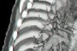
Using 3D skeletal views, orthodontists can inspect the integrity of the bone, particularly distances and periodontal defects, Mah said. An appreciation of bone levels is also possible with volumetric rendering, helping clinicians spot bone cysts or salivary gland defections and depressions.
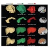
With 3D software, it's also possible to create more realistic models of patients, including soft-tissue analysis and texture maps, he pointed out.
Alveolar ridge
Volumetric imaging can clarify discrepancies in alveolar ridge shape and volume. Three-dimensional modeling can also help orthodontists select arch wires and forms in advance. These 3D radiomodels would replace plaster molds, resulting in more accurate measurements, which can be difficult to obtain when teeth are missing, Mah said.
Other 3D imaging advantages in the alveolar ridge include different views for evaluating dentition and registering the dentition to skeletal structure, including joint position.
TMJ, sinus, and airways
In addition, volumetric imaging can aid in assessing degenerative disease in the temporomandibular joint (TMJ). In terms of the sinuses and airways, advanced techniques can show potential connections between orthodontic problems and sinusitis or nasal polyps.
Looking into the future
Mah also touched on the future applications for volumetric imaging such as 3D morphometric analysis and treatment simulation. Other possibilities include registration tools for creating a therapeutic model and then taking a look into the future to theorize on potential outcomes.
"It's fairly easy to image the patient," Mah said. "It's what happens after that's important. That's where the software plays a key role. We have to ask ourselves, what additional information can be obtained with 3D? We should be using the information for more than just looking at the patient."
By Shalmali Pal
AuntMinnie.com staff writer
July 4, 2005
Related Reading
3D diagnosis and planning bring orthodontists closer to 'anatomic truth', June 2, 2005
Orthodontist advises practice needs assessment before investing in DR, May 25, 2005
MDCT advances oral and maxillofacial surgery practice, November 22, 2004
MRI clicks with TMJ diagnosis and treatment, October 15, 2004
Copyright © 2005 AuntMinnie.com