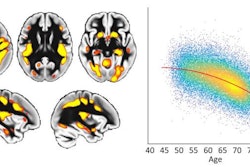
A new bioluminescence imaging technique appears to show movement of oxygen in the brain, according to research at the University of Rochester Medical Center.
The imaging technique studied in mice involves using a virus to deliver instructions to neurological cells called astrocytes and injecting a substrate called furimazine into the brain to generate light. While existing oxygen monitoring techniques provide information about a very small area of the brain, the researchers were able to observe, in real-time, a large section of the cortex of the mice.
The research is important because it opens doors for studying diseases associated with hypoxia in the brain, including Alzheimer’s, vascular dementia, and long COVID, and how a sedentary lifestyle, aging, hypertension, and other factors contribute to these diseases.
“This research demonstrates that we can monitor changes in oxygen concentration continuously and in a wide area of the brain,” said Maiken Nedergaard, MD, co-director of the Center for Translational Neuromedicine, which is based at both the University of Rochester and the University of Copenhagen. “This provides us with a more detailed picture of what is occurring in the brain in real-time, allowing us to identify previously undetected areas of temporary hypoxia, which reflect changes in blood flow that can trigger neurological deficits.”
The article highlighted by the University of Rochester Medical Center was published in Science.