Using a patient-centric temporal data model, ABM provides a visual summary of all pertinent information for breast imaging patients, enabling radiologists to rapidly assimilate this information and improve their preparation for breast imaging interpretation, according to presenter Jafi Lipson, MD.
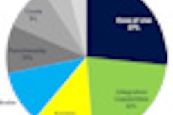
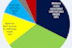
Data are extracted from the electronic medical record (EMR) and RIS; the tool brings together demographics, breast cancer risk factors, and exam indication, and it provides annotated images of both breasts. It also summarizes breast findings in a graphical form, according to the researchers from Stanford, CA.
In testing, the researchers found that ABM allows the radiologist to review and understand the same information in significantly less time than it would have taken without ABM.
In its current iteration, ABM can't be used in clinical practice. In the future, however, the researchers envision that ABM will be populated automatically and linked to the source patient data in the EMR, RIS, and PACS.
"We are currently developing the informatics infrastructure to enable linking ABM to the source clinical systems," Lipson told AuntMinnie.com. "Our vision is for ABM to be a 'living visual summary record' of breast patient information, continually updating automatically from clinical information systems as patients receive their care."