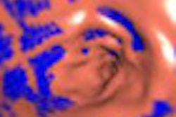
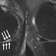
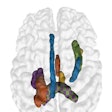
The use of high-resolution imaging modalities such as MDCT continues to increase, placing more demands on radiologists to read and interpret large numbers of images. At the same time, the accumulation of these images in PACS databases offers an opportunity for data mining to assist current patient management, said presenter Albert Montillo, PhD, who was a visiting scientist at Microsoft during the research project.
With that in mind, the research team developed an automated algorithm for segmenting anatomy of large field-of-view CT scans. Testing has yielded a high level of accuracy, according to the researchers.
The technology could be applied for applications such as automatic estimation of organ volume and density, semantic image registration for quantification of difference images, organ-driven image navigation for rapid visualization of the structure of interest, and automatic indexing of patient scans to aid in search and retrieval, Montillo said.
