MALTOMA:
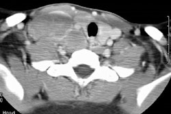
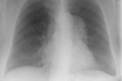
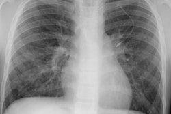
As is typical, the lesion in this case presents as a solitary pulmonary nodule within the right lower lung (it overlies the right anterior 5th rib on the PA exam, but is better seen on the lateral view). The lesion is well circumscribed and is non-calcified on computed tomography. There was no evidence of hilar or mediastinal adenopathy.


