- J Comput Assist Tomogr 1998 Jul-Aug;22(4):601-4
- Evaluation of patients with round atelectasis using
2-[18F]-fluoro-2-deoxy-D-glucose PET.
McAdams HP, Erasums JJ, Patz EF, Goodman PC, Coleman RE
Department of Radiology, Duke University Medical Center, Durham, NC 27710, USA.
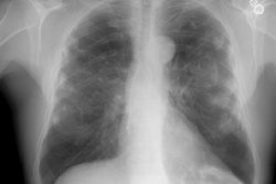
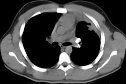
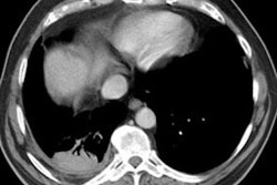
PURPOSE: Our goal was to determine the spectrum of 2-[18F]fluoro-2-deoxy-D-glucose (FDG) PET findings in patients with round atelectasis (RA). METHOD: All patients from 1992 to 1997 with radiologic features of RA and FDG-PET scans were evaluated. There were nine men ranging in age from 52 to 75 years (mean 65 years). All had chest radiographs and CT scans that were correlated with FDG-PET. FDG-PET was considered positive if lesion activity was greater than mediastinal activity and negative if lesion activity was the same as or less than mediastinal activity. RESULTS: Nine patients had 10 lesions, ranging in size from 1.2 to 5.0 cm (mean 3.1 cm). Lesion locations were right lower lobe (n = 5), left lower lobe (n = 4), and lingula (n = 1). All lesions were homogeneous and of soft tissue attenuation on CT. None contained air bronchograms or calcification. All had in-curving vessels and bronchi (comet tail sign), adjacent pleural thickening, and volume loss on CT. All lesions were negative on FDG-PET. Four lesions were percutaneously biopsied and showed chronic inflammation consistent with RA. Two lesions were unchanged on 2 and 3 year follow-up CT and were presumed to be RA as were four other lesions with characteristic CT features and negative FDG-PET. CONCLUSION: Our experience suggest that RA in not metabolically active on FDG-PET imaging. Thus, FDG-PET scans can play a role in differentiating RA from malignancy when there are few or atypical features of RA on chest radiographs and CT.
PMID: 9676452, UI: 98341206
Inhale > Asbestos
Latest in Inhalation-Lungs
Inhale > E-cigaretteVaping Associated Lung Injury
April 14, 2021

Sponsored
Inhale > Giant cell interstitial pneumonia
April 2, 2002
Inhale > Asbestos
April 2, 2002