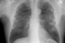
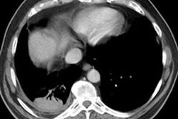
Rounded atelectasis in a patient with prior granulomatous infection
Asbestos related pleural disease is not the only cause of rounded atelectasis. The patient shown in the images below had a prior granulomatous infection with densely calcified left hilar adenopathy. There is extensive plerual thickening seen along the left anterior pleural surface. Rounded consolidated lung can be seen to abut this area of pleural thickening. Lung markings "swirl" into the area of abnormality. There is shift of the mediastinal structures to the left consistent with volume loss.