VIENNA - Children with cystic fibrosis may suffer from an unusual form of pancreatic alteration characterized by macrocysts of different sizes distributed all over the gland. Researchers from the department of pediatric surgery at Children’s Hospital La Paz in Madrid analyzed the radiologic features of pancreatic cysts in a study presented at the European Congress of Radiology on Monday.
"Cystic fibrosis is the most lethal autosomal recessive disease," said Dr. Susana Novo, who presented the study. "Pancreatic alteration in patients with cystic fibrosis is variable and seldom macroscopically seen." This complication may manifest as a complete replacement of the pancreas with fibrofatty tissue, or the replacement of the pancreas with multiple cysts, she added.
In this study sample, pancreatic ultrasound exams of 194 patients who had been diagnosed with cystic fibrosis were first reviewed. Six of these patients, ages 12 to 16 years, showed macroscopic cystic lesions in the pancreas.
CT and MR imaging followed the ultrasound exams and were used to evaluate the number, size, shape, distribution, and characteristics of the lesions.
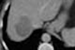
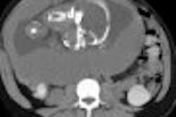
All patients showed an enlarged pancreas. However, only two patients had complete cystic transformation of the gland, with cyst sizes ranging from 2 mm to 2 cm. One patient had a rounded, well-defined, fluid-filled lesion in the tail of the pancreas. Three patients showed various multiloculated cystic lesions of different sizes, which were surrounded by normal pancreatic tissue.
On sonography, all masses were thin-walled, sonolucent masses, Novo said. On axial CT, the cysts were well outlined and had a water-like density, she added. In addition, they showed a low attenuation and homogeneous texture.
MRI demonstrated a high signal on the T2-weighted, fast spin-echo images, and a marked hypointense signal in the pancreatic tissue on the T1-weighted, spin-echo axial images.
CT and MRI were able to provide valuable information about the nature and content of the cysts, and helped to differentiate this condition from other pancreatic cystic lesions, Novo concluded.
A session attendee asked if Novo had found any correlation between the imaging findings and pancreatic patients. Because all of the patients were asymptomatic, functionality was normal, Novo said.
By Shalmali Pal
AuntMinnie.com staff writer
March 5, 2001
Click here to post your comments about this story. Please include the headline of the article in your message.
Copyright © 2001 AuntMinnie.com