Focused contrast-enhanced CT is a safe and effective way to image pregnant women for suspected appendicitis, according to the 11-year experience of radiologists at Massachusetts General Hospital in Boston.
"There are two main forms for evaluating suspected appendicitis. The first is sonography, but it remains operator-dependent and has a reported sensitivity between 75% and 90%," said Dr. Mark Mullins from MGH in a presentation at the RSNA conference on November 28.
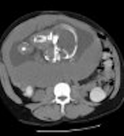
In comparison, contrast-enhanced CT had a 100% sensitivity, specificity, positive predictive value, and accuracy in his institution’s experience, Mullins said. In a retrospective evaluation, records from 1988 to 1999 were searched for the results of exams performed on pregnant women with possible appendicitis. Patients without known intrauterine pregnancies were excluded. The gestational age of the fetus was between five and 39 weeks. The most common complaint was abdominal pain, Mullins said.
From that search, 34 gravid women who had undergone CT, ultrasound, or both were identified. Results were correlated with surgical pathology or follow-up exams. The rectal contrast agent used was Gastrografin diluted in 1,000 cc of saline. One patient received the intravenous contrast Oxilan, and one other patient received Gastrografin as an oral contrast.
"We found that focused CT with contrast was well-tolerated up until the third trimester, when inability to retain the colon contrast was noted," Mullins said.
According to the results, there were three true-positive, eight true-negative, and no indeterminate CT results. In contrast, there was one false-positive, five-true negative, one true-positive, and 23 indeterminate ultrasound studies.
In terms of radiation exposure, the physicists at MGH have deemed 10 rads, or less than one rad to the fetus, as an acceptable level, Mullins said. The limited CT technique used on this patient population met those standards, he added.
In a related study, physicists from the University of Crete in Greece devised dosimetric data for performing CT in pregnant women. According to their calculations, "in CT examinations of the abdomen, doses of 30 to 43.6 mGy and of 29.1 to 42.0 mGy were measured at the measuring points in the phantom simulating pregnancy in the second and third trimesters, respectively. In CT examinations of the upper abdomen, pelvis, and thorax, both phantoms received lower doses of radiation." (Investigative Radiology, September 2000, Vol. 35:9, pp.527-522).
By Shalmali Pal
AuntMinnie.com staff writer
January 15, 2001
Click here to post your comments about this story. Please include the headline of the article in your message.
Copyright © 2001 AuntMinnie.com