Intrathoracic multicentric Castleman disease: CT findings in 12 patients.
Johkoh T, Muller NL, Ichikado K, Nishimoto N, Yoshizaki K, Honda O,
Tomiyama N, Naitoh H, Nakamura H,
Yamamoto S
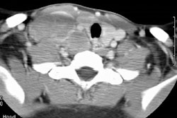
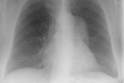
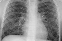
PURPOSE: To assess the computed tomographic (CT) findings of intrathoracic involvement in multicentric Castleman disease. MATERIALS AND METHODS: The study comprised 12 patients with lymph node biopsy-proved Castleman disease and multicentric involvement. The patients were aged 23-58 years (mean age, 42.9 years; five men, seven women). Seven patients underwent open lung biopsy (n = 3) or transbronchial lung biopsy (n = 4), which demonstrated lymphocytic interstitial pneumonitis. RESULTS: All patients had systemic symptoms, polyclonal hypergammaglobulinemia, and bilateral hilar and mediastinal lymph node enlargement. The nodes showed mild to moderate enhancement after intravenous administration of contrast material. At thin-section CT, all-12 patients showed poorly defined centrilobular nodules. Thin-walled cysts were present in 10 patients, thickening of the bronchovascular bundles in 10, and interlobular septal thickening in nine. Less common findings were subpleural nodules, ground-glass attenuation, air-space consolidation, and bronchiectasis. CONCLUSION: Multicentric Castleman disease is characterized by the presence of systemic symptoms, bilateral hilar and mediastinal lymphadenopathy, and centrilobular nodular opacities. The pulmonary parenchymal findings are due to the associated lymphocytic interstitial pneumonitis.