Marks MJ, Haney PJ, McDermott MP, White CS, Vennos AD
.
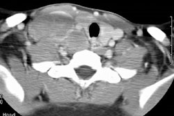
Knowledge of common and uncommon thoracic pathologic conditions in
children with acquired immunodeficiency syndrome (AIDS) can expedite disease
management. Chest radiography, computed tomography (CT), and magnetic resonance
(MR) imaging are useful in cases involving possible complications of thoracic
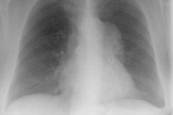
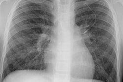
AIDS. Lymphocytic interstitial pneumonitis (LIP) is generally seen on plain
radiographs and CT scans as a diffuse, symmetric, reticulonodular or nodular
pattern, occasionally associated with mediastinal or hilar adenopathy.
Chronic consolidations and bronchiectasis may be observed in pediatric
AIDS patients with no evidence of previous LIP. Bacterial pneumonia, a
frequent initial manifestation of AIDS, appears as lobar or segmental consolidations
on radiographs. Radiographic findings of Pneumocystis carinii pneumonia,
the most common infection, include rapidly progressive increased air-space
opacity with air bronchograms. Lymphoma often appears as a mediastinal
or hilar mass, often without involvement of the lung parenchyma. Thoracic
smooth muscle tumors have also been observed in children with AIDS. Multilocular
thymic cysts have low attenuation on CT scans and increased signal intensity
on T2-weighted MR images. Most pediatric AIDS patients with cardiac disease
have cardiomegaly, often associated with pulmonary edema, at chest radiography.
An esophagogram may show ulceration, plaque formation, mucosaledema, and
dysmotility in patients with candidal esophagitis.
Lymph > LIP
Radiographics 1996 Nov;16(6):1349-1362. Thoracic disease in children
with AIDS.
Latest in Lymphoproliferative
Lymph > Nodular lymphoid hyperplasia
February 12, 2014

Sponsored
Register today for a FREE webinar May 8 at 12 Noon EDT
April 24, 2024
Lymph > Pulmonary lymphangiectasis
January 3, 2010
Lymph > General
April 2, 2002