The RSNA has launched a challenge competition to develop artificial intelligence (AI) algorithms to detect and localize cervical spine (C-spine) injuries.
The RSNA's Cervical Spine Fracture AI Challenge is designed to explore whether AI can aid in detecting and localizing cervical spine injuries. The society is hosting the challenge in collaboration with the American Society of Neuroradiology (ASNR) and the American Society of Spine Radiology (ASSR).
The challenge is being conducted on a platform provided by the data science company Kaggle and is open to everyone. The competition phase will finish in October, with the top 10 performing competitors to be awarded a total of $30,000.
Contestants will try to develop machine-learning models that match the performance of radiologists in detecting and localizing fractures within the seven vertebrae that comprise the cervical spine.
The planning task force for the challenge collected imaging data sourced from 12 sites on six continents to create its ground truth dataset. This includes more than 1,400 CT exams with diagnosed cervical spine fractures, as well as an approximately equal number of negative exams. Spine radiology specialists from the ASNR and ASSR also provided expert image-level annotations to indicate the presence, vertebral level, and location of any cervical spine fractures.
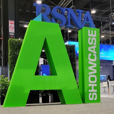
Winners will be recognized in the AI Showcase during RSNA 2022, scheduled for November 27 to December 1 in Chicago.
For the RSNA's 2021 AI challenge, over 1,500 teams registered and competed in a competition that focused on AI for neuroradiology tasks.



![A normal mammogram confirmed by three-year radiologic follow-up illustrates reader-marked regions of interest (ROIs) during (A) unaided (round 1) and (B) artificial intelligence (AI)–assisted (round 2) reading. Each colored dot represents an ROI for recall by a human reader. Readers could mark more than one ROI per case, represented by multiple dots of the same color. During AI-assisted reading, the AI system displayed three visible prompts: two with suspicion of malignancy scores of 35% (left mediolateral oblique [L MLO] and craniocaudal [L CC]) and one with a suspicion of malignancy score of 10% (right craniocaudal [R CC]), shown as polygonal overlays. Without AI, six of 10 readers (60%) marked a false-positive ROI. With AI assistance, this fell to two of 10 (20%). R MLO = right mediolateral oblique.](https://img.auntminnie.com/mindful/smg/workspaces/default/uploads/2026/07/2026-07-14-radiology-mammogram-ai-auto-bias.H0bYO8QlWs.jpg?auto=format%2Ccompress&fit=crop&h=100&q=70&w=100)


![A normal mammogram confirmed by three-year radiologic follow-up illustrates reader-marked regions of interest (ROIs) during (A) unaided (round 1) and (B) artificial intelligence (AI)–assisted (round 2) reading. Each colored dot represents an ROI for recall by a human reader. Readers could mark more than one ROI per case, represented by multiple dots of the same color. During AI-assisted reading, the AI system displayed three visible prompts: two with suspicion of malignancy scores of 35% (left mediolateral oblique [L MLO] and craniocaudal [L CC]) and one with a suspicion of malignancy score of 10% (right craniocaudal [R CC]), shown as polygonal overlays. Without AI, six of 10 readers (60%) marked a false-positive ROI. With AI assistance, this fell to two of 10 (20%). R MLO = right mediolateral oblique.](https://img.auntminnie.com/mindful/smg/workspaces/default/uploads/2026/07/2026-07-14-radiology-mammogram-ai-auto-bias.H0bYO8QlWs.jpg?auto=format%2Ccompress&fit=crop&h=112&q=70&w=112)